Play all audios:
MAIN Sir, Chicken pox is a common childhood disease caused by the varicella zoster virus. Although most cases of varicella infection resolve without any sequelae, complications have been
described.1,2 To our knowledge, this is the first case report of an orbital vasculitis following chicken pox infection in an immunocompetent patient. CASE REPORT A 7-year-old Filipino girl
presented to the paediatric department with a ‘swollen right eye’, 6 days following chicken pox infection. On examination, she was systemically unwell and pyrexial. There was a right-sided
axial proptosis with conjunctival chemosis and generalized restriction of extraocular movements. There was no relative afferent pupillary defect (RAPD) and her fundi looked healthy. She had
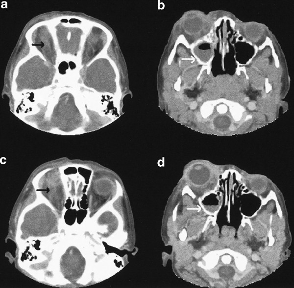
neutrophilia and the C-reactive protein (CRP) level was raised. An urgent contrast-enhanced CT scan showed inflammatory changes within the superior retro-orbital fat and opacification of the
right maxillary sinus (Figure 1a and b). The other paranasal sinuses were clear. A diagnosis of bacterial orbital cellulitis was made and treatment with intravenous benzylpenicillin and
flucloxacillin was commenced. After 4 days, she was referred to the ophthalmic adnexal service for management as the proptosis had worsened (Figure 2a). The globe was displaced downwards and
there was a mild right RAPD with mydriasis of the right pupil. Fundal examination remained unremarkable. Systemically, she was getting better with improvement in her neutrophilia and CRP
levels. An urgent repeat contrast-enhanced CT scan revealed prominent orbital blood vessels with surrounding extensive inflammatory changes. The maxillary sinusitis had improved with no
evidence of an abscess or cavernous sinus thrombosis in the right orbit (Figure 1c and d). A noninfective inflammatory orbital disease affecting the orbital vasculature was suspected and
high-dose oral prednisolone (2 mg/kg) was commenced under the cover of intravenous acyclovir (250 mg/m2 t.i.d.). There was a rapid improvement in her orbital inflammation and corresponding
signs, and the acyclovir was discontinued 2 days later. The steroid dosage was tapered off over 2 months. On review 1 month after presentation, there was complete resolution of her orbital
inflammation without any complications (Figure 2b). COMMENT This case illustrates the potential diagnostic pitfalls that can occur in the management of a ‘hot’ orbit. The clinical
presentation was suggestive of bacterial orbital cellulitis with subsequent abscess formation causing globe displacement. However, it is uncommon to develop bacterial orbital infection from
maxillary sinus disease with an intact orbital floor. The repeat CT scan did not confirm the presence of an abscess or cavernous sinus thrombosis as potential causes for the worsening
proptosis. Instead, the orbital blood vessels were found to be distended with marked surrounding inflammatory changes. The systemic improvement of the patient in the face of worsening
orbital inflammation led us to suspect the possibility of a noninfective orbital inflammatory disorder affecting the orbital vasculature. The diagnosis was supported by the rapid response to
high-dose systemic steroids. Unfortunately, a magnetic resonance angiogram that may have helped confirm the diagnosis of vasculitis was not performed.3 Varicella-associated vasculopathy is
a complication of chicken pox infection with potentially serious consequences. This has been described in the central nervous system, kidney, and retina. The pathology ranges from small
vessel vasculitis with lymphocytic infiltration of the vessel wall to giant cell arteritis involving the larger blood vessels.1,4,5,6 Fortunately, our patient experienced no permanent
sequelae from the vasculitis. In conclusion, this case demonstrates that orbital vasculitis can occur following chicken pox infection. A high index of suspicion is needed to make the correct
diagnosis as orbital vasculitis can mimic orbital cellulitis. Treatment with high-dose systemic steroids results in a rapid resolution of the inflammation. REFERENCES * Takeoka M, Takahashi
T . Infectious and inflammatory disorders of the circulatory system and stroke in childhood. _Curr Opin Neurol_ 2002; 15: 159–164. Article Google Scholar * Kuo YH, Yip Y, Chen SN .
Retinal vasculitis associated with chickenpox. _Am J Ophthalmol_ 2001; 132: 584–585. Article CAS Google Scholar * Kramer LA, Villar-Cordova C, Wheless JW, Slopis J, Yeakley J . Magnetic
resonance angiography of primary varicella vasculitis: report of two cases. _J Magn Reson Imaging_ 1999; 9: 491–496. Article CAS Google Scholar * Hayman M, Hendson G, Poskitt KJ, Conolly
M . Postvaricella angiopathy: report of a case with pathologic correlation. _Pediatr Neurol_ 2001; 24: 387–389. Article CAS Google Scholar * Caruso JM, Tung GA, Brown WD . Central nervous
system and renal vasculitis associated with primary varicella infection in a child. _Pediatrics_ 2001; 107: E9. Article CAS Google Scholar * Kleinschmidt-DeMasters BK, Gilden DH .
Varicella-Zoster virus infections of the nervous system: clinical and pathologic correlates. _Arch Pathol Lab Med_ 2001; 125: 770–780. CAS PubMed Google Scholar Download references AUTHOR
INFORMATION AUTHORS AND AFFILIATIONS * Opthalmology Department, Norfolk and Norwich University Hospital, Colney Lane, Norwich, NR4 7UZ, UK A Ang, G Galloway, M B Kashkouli & B Beigi
Authors * A Ang View author publications You can also search for this author inPubMed Google Scholar * G Galloway View author publications You can also search for this author inPubMed Google
Scholar * M B Kashkouli View author publications You can also search for this author inPubMed Google Scholar * B Beigi View author publications You can also search for this author inPubMed
Google Scholar CORRESPONDING AUTHOR Correspondence to B Beigi. RIGHTS AND PERMISSIONS Reprints and permissions ABOUT THIS ARTICLE CITE THIS ARTICLE Ang, A., Galloway, G., Kashkouli, M. _et
al._ Orbital vasculitis following varicella. A case report. _Eye_ 18, 432–433 (2004). https://doi.org/10.1038/sj.eye.6700678 Download citation * Published: 07 April 2004 * Issue Date: 01
April 2004 * DOI: https://doi.org/10.1038/sj.eye.6700678 SHARE THIS ARTICLE Anyone you share the following link with will be able to read this content: Get shareable link Sorry, a shareable
link is not currently available for this article. Copy to clipboard Provided by the Springer Nature SharedIt content-sharing initiative