Play all audios:
MAIN Sir, Primary antiphospholipid syndrome is characterized by the production of moderate to high levels of antiphospholipid antibodies, associated with thrombotic phenomena (arterial or
venous) and recurrent spontaneous abortion due to placental vascular insufficiency, in the absence of any other recognizable autoimmune disease.1 Primary antiphospholipid syndrome has been
found to be associated with ocular ischaemia and retinal vascular occlusion, due to arteriolar or venular thrombosis.2,3,4,5,6 We report a case of a 60-year-old woman with primary
antiphospholipid syndrome, who developed consecutive occlusion of the central retinal vein and ophthalmic artery. This is the first reported case in patients with this syndrome. CASE REPORT
A 60-year-old woman presented with a 1-day history of slurred speech and left-sided weakness. She also described an episode of transient dimming of vision in the right eye that lasted 4 h.
She had a history of mild hypertension and noninsulin-dependent diabetes mellitus that was diagnosed 6 months before, both of which were adequately controlled by lifestyle modification and
dietary measures alone. Neurological examination revealed a left-sided haemiparesis and a left upper motor neuron facial nerve palsy. Visual acuity was 6/7.5 OU. Funduscopic examination was
normal. Magnetic resonance imaging of the brain demonstrated the presence of a right corona radiata infarct. Duplex carotid ultrasonography showed moderate dampening of the velocity and
waveform of the right internal carotid artery blood flow compared to the left. The patient was started on ticlopidine. After 5 months, she presented with a sudden reduction in vision acuity
in the right eye to 6/18. Intraocular pressures were normal. Fundus examination revealed a right central retinal vein occlusion. She was screened for autoimmune and procoagulant disorders.
Pertinent laboratory investigations were as follows: the erythrocyte sedimentation rate was 57 mm/h; IgG anticardiolipin was raised to 33 GPL units/ml; antinuclear antibody and lupus
anticoagulants were negative. Serum total complement, C3, C4, and protein S, protein C and antithrombin III were normal. The diagnosis of primary antiphospholipid syndrome was established
and the patient was started on anticoagulation with warfarin. After 1 month, she presented with sudden deterioration of her right vision to perception of light only. A right relative
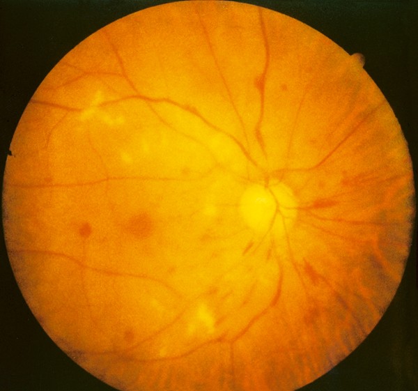
afferent pupillary defect was present. There was neovascularization of the iris, with a raised intraocular pressure of 26 mmHg. Fundus examination revealed the presence of a right ophthalmic
artery occlusion, with severe whitening of the retina and markedly attenuated retinal arterioles and venules (Figure 1). Fundus fluorescein angiography demonstrated blocked choroidal and
retinal artery filling. Transcranial doppler ultrasound of the ophthalmic arteries revealed an absent signal over the right ophthalmic artery and a normal signal from the left. The
international normalized ratio (INR) was 1.6. Immediate panretinal photocoagulation was instituted. She was started on oral acetazolamide 250 mg bid and topical timolol 0.5% bid and atropine
1% tid to the affected eye. Warfarin therapy was optimized to INR 2.5–3.0. There was gradual resolution of the iris neovascularization and eventual normalization of the intraocular
pressure. However, the final visual acuity in the right eye was hand movements only. She remained asymptomatic in the left eye. COMMENT Patients with primary antiphospholipid syndrome are at
risk of developing systemic and cerebral thromboembolism. The frequency of ocular vaso-occlusive disorders in patients with this syndrome ranges from 0.5 to 8%, with the majority affecting
the retinal vasculature.2,3,4,5 Both the arterial and venous systems may be involved. Clinical features include microaneurysms, vitreous and preretinal haemorrhage, anterior ischaemic optic
neuropathy, as well as retinal and choroidal vascular occlusions.2,3,4,5 Iris neovascularization tends to occur early,6 as was demonstrated in our patient. Castanon _et al_3 reported a high
prevalence of ocular disease in patients with primary antiphospholipid syndrome, with 88% of patients (15 out of 17 patients) demonstrating funduscopic abnormalities, and 29% of patients
having vaso-occlusive retinopathy. Consecutive central retinal vein and ophthalmic artery occlusion is extremely rare in patients with primary antiphospholipid syndrome. This is the first
reported case of this occurrence. Antiphospholipid antibodies are believed to promote thrombotic events due to its action on the phospholipid component of platelet membranes and vascular
endothelium, as well as on thrombotic factors such as local prostacyclin, antithrombin III, and protein C activation.6,7 Anticoagulation with warfarin has been advocated in patients with
vaso-occlusive retinopathy as it may assist in the reperfusion of the ischaemic retina and reduce the risk of neovascularization.8 Ophthalmologists should consider screening for
antiphospholipid antibodies in patients without obvious cardiovascular risk factors, who present with ocular and systemic vaso-occlusive disease. REFERENCES * Bolling JP, Brown GC . The
antiphospholipid antibody syndrome. _Curr Opin Ophthalmol_ 2000; 11(3): 211–213. Article CAS Google Scholar * Demirci FY, Kucukkaya R, Akarcay K, Kir N, Atamer T, Demirci H _et al_.
Ocular involvement in primary antiphospholipid syndrome. _Int Ophthalmol_ 1999; 22(6): 323–329. Article Google Scholar * Castanon C, Amigo MC, Banales JL, Nava A, Reyes PA . Ocular
vaso-occlusive disease in primary antiphospholipid syndrome. _Ophthalmology_ 1995; 102(2): 256–262. Article CAS Google Scholar * Wiechens B, Schroder JO, Potzsch B, Rochels R . Primary
antiphospholipid antibody syndrome and retinal occlusive vasculopathy. _Am J Ophthalmol_ 1997; 123(6): 848–850. Article CAS Google Scholar * Boets EP, Chaar CG, Roday K, Keunen JE,
Breedveld FC . Chorioretinopathy in primary antiphospholipid syndrome: a case report. _Retina_ 1998; 18(4): 382–385. Article CAS Google Scholar * Kleiner RC, Najarian LV, Schatter S, Jabs
DA, Patz A, Kaplan HJ . Vaso-occlusion retinopathy associated with anti-phospholipid antibodies (lupus anticoagulant retinopathy). _Ophthalmology_ 1989; 96: 896–904. Article CAS Google
Scholar * Bernard AG, Bayani N, Chretien P, Cochard C, Lelong F, Coscas G . Antiphospholipid antibodies in retinal vascular occlusions: a prospective study of 75 patients. _Arch Ophthalmol_
1994; 112: 790–795. Article Google Scholar * Kent D, Hickey-Dwyer M, Clark D . Long-term follow-up of ischaemic retinopathy in the antiphospholipid syndrome with lupus-like disease. _Eye_
2000; 14(3A): 313–317. Article Google Scholar Download references AUTHOR INFORMATION Author notes * The authors have no commercial interest in any of the material discussed in the paper.
AUTHORS AND AFFILIATIONS * Singapore National Eye Centre, 11 Third Hospital Avenue, 168751, Singapore, Singapore L Pek-Kiang Ang * Tan Tock Seng Hospital, Moulmein Road, 308433, Singapore,
Singapore A Tock-Han LIM & E-Y Yap Authors * L Pek-Kiang Ang View author publications You can also search for this author inPubMed Google Scholar * A Tock-Han LIM View author
publications You can also search for this author inPubMed Google Scholar * E-Y Yap View author publications You can also search for this author inPubMed Google Scholar CORRESPONDING AUTHOR
Correspondence to L Pek-Kiang Ang. RIGHTS AND PERMISSIONS Reprints and permissions ABOUT THIS ARTICLE CITE THIS ARTICLE Pek-Kiang Ang, L., Tock-Han LIM, A. & Yap, EY. Central retinal
vein and ophthalmic artery occlusion in primary antiphospholipid syndrome. _Eye_ 18, 439–440 (2004). https://doi.org/10.1038/sj.eye.6700685 Download citation * Published: 07 April 2004 *
Issue Date: 01 April 2004 * DOI: https://doi.org/10.1038/sj.eye.6700685 SHARE THIS ARTICLE Anyone you share the following link with will be able to read this content: Get shareable link
Sorry, a shareable link is not currently available for this article. Copy to clipboard Provided by the Springer Nature SharedIt content-sharing initiative