Play all audios:
Sir, A 24-year-old black male with sickle cell disease (SCD) ‘SS’ presented to the emergency room (ER) in sickle cell crisis with acute painless loss of vision OD, chest, and leg pains since
one day. His visual acuity was HM OD and J1 OS with 3+ afferent pupillary defect OD. Anterior segment examination was unremarkable. Ophthalmoscopy OD revealed retinal pallor with dilated
tortuous vessels. OS fundus was normal. Systemic evaluation revealed icterus with normal pulmonary, cardiovascular, or neurological examination with no history of comorbid conditions. The
haemoglobin was 9.6 gm/dl and haematocrit was 27.9%. Exchange transfusion was performed subsequent to hydration. At 1 week the visual acuity (VA) was HM OD and 20/25 OS. Ophthalmoscopy OD
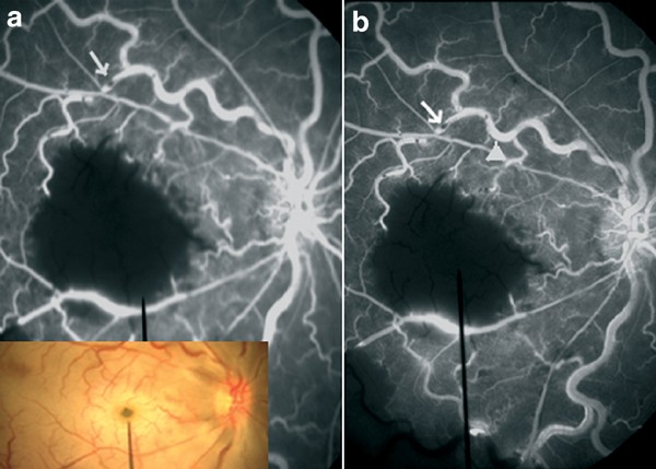
revealed pale, white, thickened retinal lesion centred on the fovea, arterial attenuation, cherry red spot, and pale optic disc (Figure 1 inset). Fluorescein angiography (FA) OD demonstrated
perifoveal arteriole occlusions with enlargement of the FAZ (Figure 1). Vessels had areas of blocked fluorescein in early and late phases (Figure 1a and b), while hyperfluorescein areas
(Figure 1b) in the late phases (staining). FA OS was normal. At 3 months VA was CF 3 feet OD, 20/20 OS. Ophthalmoscopy OD revealed pale disc with marked arterial attenuation and pigmentary
changes in the macula (Figure 2a). Macular perimetry (Nidek Technologies, Vigonza, Italy) revealed unstable fixation with absolute macular scotoma OD with normal findings OS (Figure 2b-d).
His haemoglobin was 9.5 gm/dl and haematocrit was 26.0%. COMMENTS Occlusive disease of the peri-foveal arterioles is known to occur in sickle cell disease.1, 2 We present a dramatic
occlusive event of the macula surrounding the foveal avascular zone, resulting in severe and permanent loss of vision. We are unaware of any previous report of FA showing possible
microemboli in the retinal vessels with macular infarction in SCD. Direct sickling may cause occlusion in arterioles or in capillary beds creating a ‘log-jam’ in the arterioles.3 In this
case, the occlusions surrounded the macula from multiple directions, suggesting perifoveal arteriolar occlusion due to microemboli and/or slugging of sickle cells with slowed circulation in
the venules. Our patient presented with an acute visual loss resulting in irreversible macular infarction. His visual recovery was minimal because of delay in presentation. REFERENCES *
Acacio I, Goldberg MF . Peripapillary and macular vessel occlusions in sickle cell anemia. _Am J Ophthalmol_ 1973; 75: 861–866. Article CAS Google Scholar * Welch RB, Goldberg MF .
Sickle-cell hemoglobin and its relation to fundus abnormality. _Arch Ophthalmol_ 1966; 75: 353–362. Article CAS Google Scholar * Goldberg MF, Galinos S, Lee CB, Stevens T, Woolf MB .
Editorial: Macular ischemia and infarction in sickling. _Invest Ophthalmol._ 1973; 12: 633–635. CAS PubMed Google Scholar Download references AUTHOR INFORMATION AUTHORS AND AFFILIATIONS *
Department of Ophthalmology, University of Florida, College of Medicine, 580 W 8th street, Jacksonville, 32209, FL, USA K V Chalam & V A Shah Authors * K V Chalam View author
publications You can also search for this author inPubMed Google Scholar * V A Shah View author publications You can also search for this author inPubMed Google Scholar CORRESPONDING AUTHOR
Correspondence to K V Chalam. ADDITIONAL INFORMATION Proprietary Interest: None. Presented as a poster at the 21st Annual Meeting of the American Society of Retina Specialists, New York, NY,
August 2003. RIGHTS AND PERMISSIONS Reprints and permissions ABOUT THIS ARTICLE CITE THIS ARTICLE Chalam, K., Shah, V. Macular infarction a presentation of sickle cell crisis. _Eye_ 18,
1277–1278 (2004). https://doi.org/10.1038/sj.eye.6701409 Download citation * Published: 23 April 2004 * Issue Date: 01 December 2004 * DOI: https://doi.org/10.1038/sj.eye.6701409 SHARE THIS
ARTICLE Anyone you share the following link with will be able to read this content: Get shareable link Sorry, a shareable link is not currently available for this article. Copy to clipboard
Provided by the Springer Nature SharedIt content-sharing initiative