Play all audios:
Sir, The Peters' anomaly is characterized by central corneal opacity (leukoma), thinning of the posterior aspect of the cornea, iridocorneal adhesions, and keratolenticular adhesion or
cataract. The presence of lens abnormalities in Peters' anomaly is more frequently associated with systemic anomalies.1 Peters' plus syndrome (PPS) is a rare entity that combines
anterior chamber abnormality of Peters' anomaly with other systemic anomalies such as cleft lip and palate, short stature, broad hands and feet, and variable mental delay.2 The etiology
is unknown, but may involve abnormal neural crest development. Peters' anomaly has been reported to be caused by mutations in several genes such as _PAX6, PITX2, PITX3, and CYP1B1_.2
PPS follows an autosomal-recessive pattern of inheritance,2 but the causative gene of this syndrome remains unknown. Previous pathologic reports in Peters' anomaly revealed attenuation
or absence of Descemet's membrane and corneal endothelium in the central posterior cornea, with iridocorneal adhesion or keratolenticular adhesion.3, 4, 5 We report, herein, the
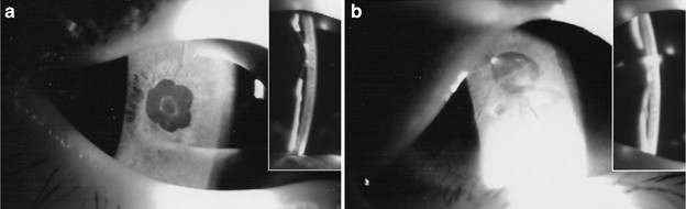
ultrastructure of anterior lens capsule in a patient with PPS. _CASE REPORT_ A 4-year-old boy was found to have bilateral cataract and referred to our department. Bilateral nystagmus was
found. We observed a poor ambulatory activity of the patient while occluding his left eye with eye patch. Slit-lamp examination showed bilateral cataract with bulging anterior lens surface
that adheres to the posterior cornea (Figure 1). Mild opacity was detected in the corneal stroma in front of the adhesion site. Microcornea and posterior synechia were also found in both
eyes. Pediatric evaluation revealed short stature (<3%), low body weight (<3%), speech, and motor developmental delay. Growth hormone deficiency was also detected by stimulation test.
The patient received extracapsular lens extraction in the right eye, with keratolenticular adhesion separated by viscoelastic material, and the anterior lens capsule removed by can-opening
capsulectomy. Aspirated lens material was sent for virus culture and Herpes virus polymerase chain reaction (PCR), with both test results came out to be negative. Anterior lens capsule was
fixed in 2% glutaraldehyde/phosphate buffer, and then washed three times in phosphate buffer. The specimen was post-fixed with 1% osmium tetraoxide. After dehydration with alcohol series, it
was embedded with epoxy/resin and sectioned. The section was examined under a JEOL electron microscope (JEM 1230). We observed villus-like processes emanated from the anterior lens capsule,
with two cells still attached to the process tips. These cells were presumed to be sloughed-off corneal endothelial cells, as the lens capsule adhered to the corneal endothelial side before
surgical separation (Figure 2, En). Intact lens epithelial cells could be found adjacent to the lens capsule (Figure 2, Ep). COMMENT Keratolenticular adhesion is characteristic in PPS.
Various pathologic results have been reported in the literature.3, 4, 5 The lens may be adherent to the corneal stroma with absence of Descemet's membrane and lens capsule.4 A
stalk-like connection between lens and cornea has been reported as well, and the Descemet's membrane was deflected over onto the stalk.5 The lens may be in contact or embedded in the
defective posterior corneal surface, but with an intact anterior lens capsule.4 The lens may be only opposed and not adherent to the posterior surface of the cornea, with both the anterior
lens capsule and cornea intact.3 To our knowledge, villus-like processes extending from the anterior lens capsular surface has never been reported in literature. These processes align
regularly along the margin, with a narrow connecting neck. These processes were about 0.5 _μ_m in width and 10–15 _μ_m in length, with some electron-dense particles between them. Since the
syndrome is genetically heterogeneous, this finding may not be generally applicable. In literature, several developmental theories have been proposed for the pathogenesis of Peters'
anomaly, and none of them explains all the clinical and histopathologic findings in all forms of Peters' anomalies.2 A failure of neural crest cell differentiation destined for corneal
endothelium and Descemet's membrane is considered the most reasonable explanation for the keratolenticular or iridocorneal adhesion. We propose that these villus-like processes might
represent the developmental remnants that fail to regress while corneal endothelium separates from the underlying lens epithelium. REFERENCES * Sabates MA, Caldwell DR, Ellis Jr GS .
Dysgenesis of the anterior segment and globe. In: Wright KW, (ed). _Pediatric Ophthalmology and Strabismus_. Mosby: St Louis, MA, 1995; 303–320. Google Scholar * Maillette de Buy
Wenniger-Prick LJ, Hennekam RC . The Peters' plus syndrome: a review. _Ann Genet_ 2002; 45: 97–103. Article Google Scholar * Schottenstein EM . Peters' anomaly. In: Ritch R,
Shields MB, Krupin T, (eds). _The Glaucomas_, 2nd ed. Mosby: St Louis, MA, 1996; 887–897. Google Scholar * Stone DL, Kenyon KR, Green WR, Ryan SJ . Congenital central corneal leukoma
(Peters' anomaly). _Am J Ophthalmol_ 1976; 81: 173–193. Article CAS Google Scholar * Harden AF, Mooney DJ . Congenital keratolenticular adhesion. _Am J Ophthalmol_ 1970; 70: 975–977.
Article CAS Google Scholar Download references AUTHOR INFORMATION AUTHORS AND AFFILIATIONS * Department of Ophthalmology, Taipei Veterans General Hospital, National Yang-Ming University
School of Medicine, Taiwan D-K Hwang, M-Y Yen & A-G Wang * Department of Pathology, Taipei Veterans General Hospital, National Yang-Ming University School of Medicine, Taiwan A-H Yang
Authors * D-K Hwang View author publications You can also search for this author inPubMed Google Scholar * A-H Yang View author publications You can also search for this author inPubMed
Google Scholar * M-Y Yen View author publications You can also search for this author inPubMed Google Scholar * A-G Wang View author publications You can also search for this author inPubMed
Google Scholar CORRESPONDING AUTHOR Correspondence to A-G Wang. RIGHTS AND PERMISSIONS Reprints and permissions ABOUT THIS ARTICLE CITE THIS ARTICLE Hwang, DK., Yang, AH., Yen, MY. _et al._
Ultrastructure of anterior lens capsule in Peters' plus syndrome. _Eye_ 21, 862–864 (2007). https://doi.org/10.1038/sj.eye.6702731 Download citation * Published: 09 February 2007 *
Issue Date: 01 June 2007 * DOI: https://doi.org/10.1038/sj.eye.6702731 SHARE THIS ARTICLE Anyone you share the following link with will be able to read this content: Get shareable link
Sorry, a shareable link is not currently available for this article. Copy to clipboard Provided by the Springer Nature SharedIt content-sharing initiative