Play all audios:
ABSTRACT PURPOSE To evaluate the prevalence and clinical features of focal choroidal excavation (FCE) in patients presenting with central serous chorioretinopathy (CSC). METHODS This is a
retrospective consecutive case series of consecutive patients with CSC who were referred for spectral domain optical coherence tomography (SD-OCT) between January 2010 and December 2011.
Medical records were reviewed and clinical features including presence of FCE in SD-OCT, fluorescence angiography (FA), and indocyanine green angiography (ICGA) were studied. RESULTS Among
the 116 CSC patients assessed, FCE was found in 11 eyes of 7 (6.0%) patients. FCE was associated with subretinal fluid in six eyes of six patients and serous pigment epithelial detachment in
three eyes of two patients. The mean central subfield retinal thickness of CSC eyes with FCE was 283.7 _μ_m, compared with 377.5 _μ_m for CSC eyes without FCE (Mann–Whitney _U_-test,
_P_=0.020). Five FCE eyes of five patients had focal leakage on FA. Choroidal hyperpermeability on ICGA was found in seven CSC eyes with FCE, with four eyes showing hypofluorescent spot
corresponding to the FCE. After a mean follow-up of 16 months, visual acuity of all 11 eyes with FCE remained stable or improved at the last follow-up. CONCLUSION FCE is not an uncommon
feature in patients with CSC and might be associated with choroidal hemodynamic disturbances. SIMILAR CONTENT BEING VIEWED BY OTHERS EN-FACE OPTICAL COHERENCE TOMOGRAPHY HYPERREFLECTIVE FOCI
OF CHORIOCAPILLARIS IN CENTRAL SEROUS CHORIORETINOPATHY Article Open access 03 May 2023 CHOROIDAL BLOOD FLOW CHANGES IN CENTRAL SEROUS CHORIORETINOPATHY Article Open access 22 March 2025
CHOROIDAL MODIFICATIONS ASSESSED BY MEANS OF CHOROIDAL VASCULARITY INDEX AFTER ORAL EPLERENONE TREATMENT IN CHRONIC CENTRAL SEROUS CHORIORETINOPATHY Article 19 May 2022 INTRODUCTION Focal
choroidal excavation (FCE) is defined as one or more focal excavations in the choroidal layer detected by optical coherence tomography (OCT), in which the overlying retina appeared to be
normal and was first reported by Jampol _et al_ in 2006.1 Clinically, FCE appears as a small localized lesion with focal pigmentary changes in fundus examination. The exact etiology of FCE
remains unknown and is postulated to be an idiopathic congenital condition. Previous studies have described two patterns of FCE: the conforming type in which the outer retinal layers conform
to retinal pigment epithelial (RPE) alterations within the excavation; and the nonconforming type, in which there is separation between the outer retina and the RPE within the excavation.2,
3 Since the first description of FCE, FCE has been found to be associated with a number of conditions including central serious chorioretinopathy (CSC), polypoidal choroidal vasculopathy
(PCV), choroidal neovascularization (CNV), and age-related macular degeneration (AMD).2, 3, 4, 5, 6 In particular, there appeared to be a higher prevalence of FCE in patients with CSC.3, 4,
5, 6, 7 The aim of this study is to evaluate the prevalence, clinical features and outcome of FCE in patients presenting with CSC. MATERIAL AND METHODS This was a retrospective study of
consecutive cases with a clinical diagnosis of CSC confirmed by spectral domain OCT (SD-OCT) assessment in the Hong Kong Eye Hospital between January 2010 and December 2011. The study
adhered to the Declaration of Helsinki and was approved by the local Ethics Committee. The medical notes of the patents were reviewed and clinical data such as visual acuity, ophthalmoscopic
findings, SD-OCT, fundus autofluorescence (FAF) imaging, fluorescein angiography (FA), and indocyanine green angiography (ICGA) findings were recorded. SD-OCT, FA, and ICGA were performed
using the Spectralis HRA-OCT system (Heidelberg Engineering, Heidelberg, Germany). Snellen visual acuity was converted to logarithm of the minimum angle of resolution (logMAR) units for
statistical analysis. Statistical analysis was performed using a statistical software (StatPlus:Mac 2009, AnalystSoft Inc., Vancouver, BC, Canada). Demographic characteristics of the
patients were summarized by descriptive statistics. Comparisons of continuous variables were performed using Mann–Whitney _U_-test or Wilcoxon sign-ranked test. A _P_-value of <0.05 was
considered as statistically significant. RESULTS PATIENTS’ CHARACTERISTICS A total of 116 patients with a clinical diagnosis of CSC were included in the study. FCE was found in 11 eyes of 7
(6.0%) patients, including 5 males and 2 females. All patients were of Chinese ethnicity. The mean±SD age of the CSC patients with FCE was 44.4±5.9 years (range, 37–56 years), compared with
45.4±9.6 years for CSC patients without FCE (range, 22–78 years; Mann–Whitney _U_-test, _P_=0.69). The mean spherical equivalent refractive error of eyes with FCE was −1.9±2.2D (range,
−0.13D to −5.5D), compared with the mean spherical equivalent refractive error of −0.60D±1.7D (range, +2.0D to −6.6D) for eyes without FCE (Mann–Whitney _U_-test, _P_=0.063). The primary
complaints of the CSC patients with FCE at presentation included blurring of vision (five patients), metamorphopsia (one patient), and scotoma (one patient). Three (42.8%) patients had a
history of smoking and one patient had a history of intermittent oral corticosteroid use for asthma 3 years before presentation. SD-OCT AND ANGIOGRAPHY FINDINGS Four (57.1%) patients were
found to have FCE in both eyes on SD-OCT imaging. One of the seven patients had two excavations in one eye, while the remaining six patients only had one FCE lesion at the macula. The
mean±SD central subfield retinal thickness (CSRT) of eyes with FCE at initial presentation was 311.9±88 _μ_m (range, 155–454 _μ_m). This compared with the mean±SD CSRT of 377.5±145 _μ_m in
patients without FCE (range, 179–974 _μ_m). The mean CSRT of CSC eyes with FCE was significantly lower compared with those without FCE (Mann–Whitney U-test, _P_=0.020). FAF was performed in
five eyes and all showed mixture of increased and reduced autofluorescence at the site of FCE. Five eyes of five FCE patients had focal leakage on FA, four of which had ink-blot leakage
appearance and one had smokestack leakage appearance. ICGA demonstrated localized hypofluorescent spot during both early and late phases in four FCE eyes, and diffuse hyperfluorescence in
the late phase owing to choroidal hyperpermeability in seven FCE eyes. DISEASE COURSE OF FCE EYES The mean±SD follow-up duration of the FCE patients was 16.5±13.0 months (range, 1–30
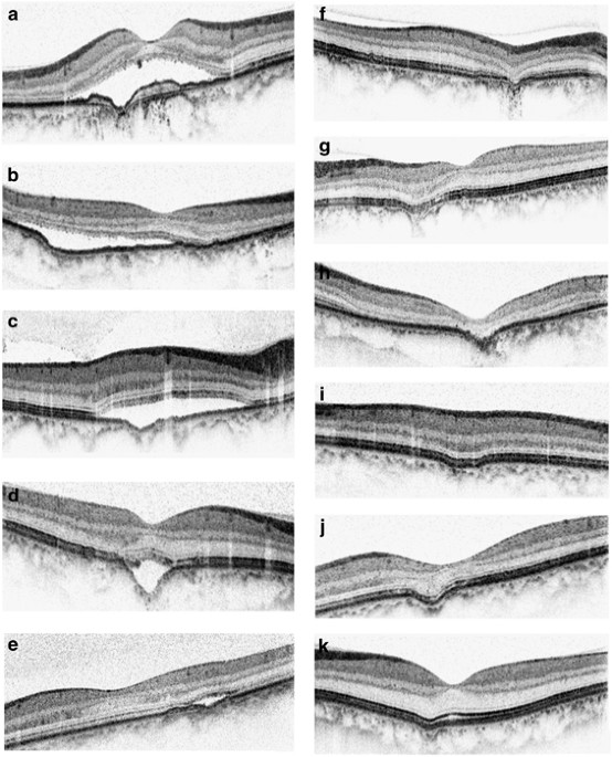
months). At baseline, the mean±SD logMAR visual acuity of the 11 eyes with FCE was 0.24±0.34 (range, 0.00–0.5). Six eyes had nonconforming FCE and five eyes had conforming FCE at the initial
presentation (Figure 1). Nine eyes with FCE were observed without any treatment. Six eyes had subretinal fluid and three eyes had serous pigment epithelial detachment at baseline. There was
no serial change in the appearance of excavation except that one case developed increase in SRF after 2 months of follow-up (Figure 2). Two eyes of nonconforming FCE with serous macular
detachment due to subretinal fluid were treated with photodynamic therapy (PDT) using half-dose (3 mg/m2) verteporfin (Visudyne, Novartis, Basel, Switzerland) and standard laser fluence (50
J/cm2). In both cases, SRF resolved completely and there were improvements in visual acuity after treatment (Figure 3). At the final follow-up visit, visual acuity of all 11 eyes with FCE
remained stable with the mean±SD logMAR visual acuity of 0.20±0.29 (range, 0.0–0.4; Wilcoxon sign-ranked test, _P_=0.27). DISCUSSION Several previous studies have reported a possible
association between FCE and CSC.3, 4, 5, 6, 7 In our study, the prevalence of FCE in CSC patients was found to be 6.0%, which was slightly lower compared with the prevalence of 7.8% reported
by Ellabban _et al_4 in 116 eyes with CSC. However, our rate was higher than the prevalence of 2.8% reported by Suzuki _et al_6 in another Japanese study of 248 eyes with CSC. However, in
contrast with previous studies, our study showed a much higher prevalence of bilateral FCE, in which four (57%) of the seven patients were found to have FCE in both eyes. This rate was
higher compared with previous studies in which 0–24% of patients had FCE bilaterally.3, 4, 5, 6, 7, 8, 9 The natural course of FCE remains unclear as most of the published studies were
cross-sectional studies. There were only a few studies with longitudinal follow-up data available and the majority of the excavations appeared to remain stable with follow-up.3, 4, 6, 10 In
our study, two eyes with FCE and active CSC were treated with PDT using half-dose verteporfin and standard laser fluence. Both patients had visual improvement with complete resolution of
subretinal fluid despite persistence of FCE after treatment. With the use of OCT and ICGA, previous studies have reported thinning of the underlying choriocapillaris in CSC eyes with FCE.4,
9, 10 This can be reflected by the presence of hypofluorescent spots that we observed in ICGA. Moreover, we also found that CSC eyes with FCE had significantly thinner CSRT compared with
those without FCE. This might be owing to less severe leakage from the choroidal circulation in CSC eyes with FCE due to loss of choriocapillaris. Choroidal hyperpermeability is one of the
main characteristic features of CSC and this can also be observed in the ICGA of all FCE eyes with CSC. Therefore, the choroidal hemodynamic disturbances in CSC eyes with FCE might involve a
mixture of diffuse choroidal hyperpermeability with localized thinning of choriocapillaris. In addition to CSC, FCE have also been shown to be associated with other retinal conditions
including PCV, AMD, and idiopathic CNV.5, 8, 9, 10, 11, 12, 13 Therefore, patients with FCE should be monitored regularly for the potential development of other retinal diseases. In
particular, CNV complexes have been reported to develop within the boundary of the excavation. Most of these CNV were of classic leakage pattern on FA and these cases usually respond well to
intravitreal antivascular endothelial growth factor therapy.12, 13 There are several limitations associated with our study including the retrospective nature and the small number of cases.
Nonetheless, we were able to follow-up these eyes and evaluated the FCE changes associated with CSC longitudinally. Our findings confirmed that FCE is not an uncommon feature in patients
with CSC and it may be associated with choroidal hemodynamic disturbances. Moreover, FCE appeared to be more commonly seen in Asian populations as the previous reports were mostly from
patients of Asian descents. Further prospective study with larger sample size will be useful to evaluate the natural history and the long-term treatment outcome of CSC eyes with FCE.
REFERENCES * Jampol LM, Shankle J, Schroeder R, Tornambe P, Spaide RF, Hee MR . Diagnostic and therapeutic challenges. _Retina_ 2006; 26: 1072–1076. Article Google Scholar * Wakabayashi Y,
Nishimura A, Higashide T, Ijiri S, Sugiyama K . Unilateral choroidal excavation in the macula detected by spectral-domain optical coherence tomography. _Acta Ophthalmol_ 2010; 88: e87–e91.
Article Google Scholar * Margolis R, Mukkamala SK, Jampoll LM, Spaide RF, Ober MD, Sorenson JA _et al_. The expanded spectrum of focal choroidal excavation. _Arch Ophthalmol_ 2011; 129:
1320–1325. Article Google Scholar * Ellabban AA, Tsujikawa A, Ooto S, Yamashiro K, Oishi A, Kakat I _et al_. Focal choroidal excavation in eyes with central serous chorioretinopathy. _Am J
Ophthalmol_ 2013; 156: 673–683. Article Google Scholar * Shinojima A, Kawamura A, Mori R, Yuzawa M . Morphologic features of focal choroidal excavation on spectral domain optical
coherence tomography with simultaneous angiography. _Retina_ 2014; 34: 1407–1414. Article Google Scholar * Suzuki M, Gomi F, Hara C, Sawa M, Nishida K . Characteristics of central serous
chorioretinopathy complicated by focal choroidal excavation. _Retina_ 2014; 34: 1216–1222. Article Google Scholar * Guo J, Zhong L, Jiang C, Zhou X, Xu G, Wang W _et al_. Clinical and
optic coherence tomography findings of focal choroidal excavation in Chinese patients. _BMC Ophthalmology_ 2014; 14: 63. Article Google Scholar * Obata R, Takahashi H, Ueta T, Yuda K, Kure
K, Yanagi Y . Tomographic and angiographic characteristics of eyes with macular focal choroidal excavation. _Retina_ 2013; 33: 1201–1210. Article Google Scholar * Lee CS, Woo SJ, Kim YK,
Hwang DJ, Kang HM, Kim H _et al_. Clinical and spectral-domain optical coherence tomography findings in patients with focal choroidal excavation. _Ophthalmology_ 2014; 121: 1029–1035.
Article Google Scholar * Lim FP, Loh BK, Cheung CM, Lim LS, Chan CM, Wong DW . Evaluation of choroidal excavation in the macula using swept- source optical coherence tomography. _Eye
(Lond)_ 2014; 28: 1088–1094. Article CAS Google Scholar * Say EA, Jani PD, Appenzeller MF, Houghton OM . Focal choroidal excavation associated with polypoidal choroidal vasculopathy.
_Ophthalmic Surg Lasers Imaging Retina_ 2013; 44: 409–411. Article Google Scholar * Lee JH, Lee WK . Choroidal neovascularization associated with focal choroidal excavation. _Am J
Ophthalmol_ 2014; 157: 710–718 e1. Article Google Scholar * Xu H, Zeng F, Shi D, Sun X, Chen X, Bai Y . Focal choroidal excavation complicated by choroidal neovascularization.
_Ophthalmology_ 2014; 121: 246–250. Article Google Scholar Download references AUTHOR INFORMATION AUTHORS AND AFFILIATIONS * Hong Kong Eye Hospital, Hong Kong SAR, China F O J Luk, A C T
Fok, A Lee & A T W Liu * Department of Ophthalmology and Visual Sciences, The Chinese University of Hong Kong, Hong Kong SAR, China F O J Luk, A C T Fok, A Lee, A T W Liu & T Y Y Lai
Authors * F O J Luk View author publications You can also search for this author inPubMed Google Scholar * A C T Fok View author publications You can also search for this author inPubMed
Google Scholar * A Lee View author publications You can also search for this author inPubMed Google Scholar * A T W Liu View author publications You can also search for this author inPubMed
Google Scholar * T Y Y Lai View author publications You can also search for this author inPubMed Google Scholar CORRESPONDING AUTHOR Correspondence to F O J Luk. ETHICS DECLARATIONS
COMPETING INTERESTS Dr Timothy Lai has received honorarium for consultancy and lecture fees from Alcon, Allergan Inc, Bausch & Lomb, Bayer Healthcare, Heidelberg Engineering and Novartis
Pharmapeuticals. The remaining authors declare no conflict of interest. ADDITIONAL INFORMATION This study was presented in part as a poster at the American Academy of Ophthalmology Annual
Meeting in November 2013 RIGHTS AND PERMISSIONS Reprints and permissions ABOUT THIS ARTICLE CITE THIS ARTICLE Luk, F., Fok, A., Lee, A. _et al._ Focal choroidal excavation in patients with
central serous chorioretinopathy. _Eye_ 29, 453–459 (2015). https://doi.org/10.1038/eye.2015.31 Download citation * Received: 06 October 2014 * Accepted: 24 December 2014 * Published: 27
March 2015 * Issue Date: April 2015 * DOI: https://doi.org/10.1038/eye.2015.31 SHARE THIS ARTICLE Anyone you share the following link with will be able to read this content: Get shareable
link Sorry, a shareable link is not currently available for this article. Copy to clipboard Provided by the Springer Nature SharedIt content-sharing initiative