Play all audios:
ABSTRACT The trichromatic primate retina parses the colour content of a visual scene into ‘red/green’ and ‘blue/yellow’ representations1,2. Cortical circuits must combine the information
encoded in these colour-opponent signals to reconstruct the full range of perceived colours3. Red/green and blue/yellow inputs are relayed by the lateral geniculate nucleus (LGN) of thalamus
to primary visual cortex (V1), so understanding how cortical circuits transform these signals requires understanding how LGN inputs to V1 are organized. Here we report direct recordings
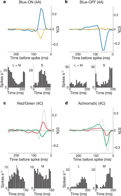
from LGN afferent axons in muscimol-inactivated V1. We found that blue/yellow afferents terminated exclusively in superficial cortical layers 3B and 4A, whereas red/green afferents were
encountered only in deeper cortex, in lower layer 4C. We also describe a distinct cortical target for ‘blue-OFF’ cells, whose afferents terminated in layer 4A and seemed patchy in
organization. The more common ‘blue-ON’ afferents were found in 4A as well as lower layer 2/3. Chromatic information is thus conveyed to V1 by parallel, anatomically segregated
colour-opponent systems, to be combined at a later stage of the colour circuit. Access through your institution Buy or subscribe This is a preview of subscription content, access via your
institution ACCESS OPTIONS Access through your institution Subscribe to this journal Receive 51 print issues and online access $199.00 per year only $3.90 per issue Learn more Buy this
article * Purchase on SpringerLink * Instant access to full article PDF Buy now Prices may be subject to local taxes which are calculated during checkout ADDITIONAL ACCESS OPTIONS: * Log in
* Learn about institutional subscriptions * Read our FAQs * Contact customer support SIMILAR CONTENT BEING VIEWED BY OTHERS CHROMATIC MICROMAPS IN PRIMARY VISUAL CORTEX Article Open access
19 April 2021 LINEAR AND NONLINEAR CHROMATIC INTEGRATION IN THE MOUSE RETINA Article Open access 26 March 2021 THE NEURAL ORIGIN FOR ASYMMETRIC CODING OF SURFACE COLOR IN THE PRIMATE VISUAL
CORTEX Article Open access 15 January 2024 REFERENCES * Lennie, P. & D'Zmura, M. Mechanisms of color vision. _Crit. Rev. Neurobiol._ 3, 333–400 (1988) CAS PubMed Google Scholar *
Dacey, D. M. Parallel pathways for spectral coding in primate retina. _Annu. Rev. Neurosci._ 23, 743–775 (2000) Article CAS Google Scholar * De Valois, R. L., Cottaris, N. P., Elfar, S.
D., Mahon, L. E. & Wilson, J. A. Some transformations of color information from lateral geniculate nucleus to striate cortex. _Proc. Natl Acad. Sci. USA_ 97, 4997–5002 (2000) Article
ADS CAS Google Scholar * Dacey, D. M., Peterson, B. B., Robinson, F. R. & Gamlin, P. D. Fireworks in the primate retina. _In vitro_ photodynamics reveals diverse LGN-projecting
ganglion cell types. _Neuron_ 37, 15–27 (2003) Article CAS Google Scholar * Leventhal, A. G., Rodieck, R. W. & Dreher, B. Retinal ganglion cell classes in the Old World monkey:
Morphology and central projections. _Science_ 213, 1139–1142 (1981) Article ADS CAS Google Scholar * Michael, C. R. Retinal afferent arborization patterns, dendritic field orientations,
and the segregation of function in the lateral geniculate nucleus of the monkey. _Proc. Natl Acad. Sci. USA_ 85, 4914–4918 (1988) Article ADS CAS Google Scholar * Conley, M. &
Fitzpatrick, D. Morphology of retinogeniculate axons in the macaque. _Vis. Neurosci._ 2, 287–296 (1989) Article CAS Google Scholar * Hubel, D. & Wiesel, T. Laminar and columnar
distribution of geniculo-cortical fibers in the macaque monkey. _J. Comp. Neurol._ 146, 421–450 (1972) Article CAS Google Scholar * Hendrickson, A. E., Wilson, J. R. & Ogren, M. P.
The neuroanatomical organization of pathways between the dorsal lateral geniculate nucleus and visual cortex in Old World and New World primates. _J. Comp. Neurol._ 182, 123–136 (1978)
Article CAS Google Scholar * Blasdel, G. G. & Lund, J. S. Termination of afferent axons in macaque striate cortex. _J. Neurosci._ 3, 1389–1413 (1983) Article CAS Google Scholar *
Rodieck, R. W. & Watanabe, M. Survey of the morphology of macaque retinal ganglion cells that project to the pretectum, superior colliculus, and parvicellular laminae of the lateral
geniculate nucleus. _J. Comp. Neurol._ 338, 289–303 (1993) Article CAS Google Scholar * Martin, P. R., White, A. J., Goodchild, A. K., Wilder, H. D. & Sefton, A. E. Evidence that
blue-on cells are part of the third geniculocortical pathway in primates. _Eur. J. Neurosci._ 9, 1536–1541 (1997) Article CAS Google Scholar * Hendry, S. H. & Reid, R. C. The
koniocellular pathway in primate vision. _Annu. Rev. Neurosci._ 23, 127–153 (2000) Article CAS Google Scholar * Hendry, S. H. & Yoshioka, T. A neurochemically distinct third channel
in the macaque dorsal lateral geniculate nucleus. _Science_ 264, 575–577 (1994) Article ADS CAS Google Scholar * Wiesel, T. N. & Hubel, D. H. Spatial and chromatic interactions in
the lateral geniculate body of the rhesus monkey. _J. Neurophysiol._ 29, 1115–1156 (1966) Article CAS Google Scholar * Schiller, P. H. & Malpeli, J. G. Functional specificity of
lateral geniculate nucleus laminae of the rhesus monkey. _J. Neurophysiol._ 41, 788–797 (1978) Article CAS Google Scholar * Derrington, A. M., Krauskopf, J. & Lennie, P. Chromatic
mechanisms in lateral geniculate nucleus of macaque. _J. Physiol. (Lond.)_ 357, 241–265 (1984) Article CAS Google Scholar * Reid, R. C. & Shapley, R. M. Space and time maps of cone
photoreceptor signals in macaque lateral geniculate nucleus. _J. Neurosci._ 22, 6158–6175 (2002) Article CAS Google Scholar * Valberg, A., Lee, B. B. & Tigwell, D. A. Neurones with
strong inhibitory S-cone inputs in the macaque lateral geniculate nucleus. _Vision Res._ 26, 1061–1064 (1986) Article CAS Google Scholar * Ringach, D. L., Sapiro, G. & Shapley, R. A
subspace reverse-correlation technique for the study of visual neurons. _Vision Res._ 37, 2455–2464 (1997) Article CAS Google Scholar * Dacey, D. M. & Lee, B. B. The ‘blue-on’
opponent pathway in primate retina originates from a distinct bistratified ganglion cell type. _Nature_ 367, 731–735 (1994) Article ADS CAS Google Scholar * Calkins, D. J. &
Sterling, P. Evidence that circuits for spatial and color vision segregate at the first retinal synapse. _Neuron_ 24, 313–321 (1999) Article CAS Google Scholar * Ahmad, K. M., Klug, K.,
Herr, S., Sterling, P. & Schein, S. Cell density ratios in a foveal patch in macaque retina. _Vis. Neurosci._ 20, 189–209 (2003) Article Google Scholar * Klug, K., Herr, S., Ngo, I.
T., Sterling, P. & Schein, S. J. Macaque retina contains an S-cone OFF midget pathway. _J. Neurosci._ 23, 9881–9887 (2003) Article CAS Google Scholar * Blasdel, G. G. &
Fitzpatrick, D. Physiological organization of layer 4 in macaque striate cortex. _J. Neurosci._ 4, 880–895 (1984) Article CAS Google Scholar * Chapman, B., Zahs, K. R. & Stryker, M.
P. Relation of cortical cell orientation selectivity to alignment of receptive fields of the geniculocortical afferents that arborize within a single orientation column in ferret visual
cortex. _J. Neurosci._ 11, 1347–1358 (1991) Article CAS Google Scholar * Stockman, A., MacLeod, D. I. & Johnson, N. E. Spectral sensitivities of the human cones. _J. Opt. Soc. Am. A_
10, 2491–2521 (1993) Article ADS CAS Google Scholar * Wandell, B. A. _Foundations of Vision_ 413–421 (Sinauer, Sunderland, MA, 1995) Google Scholar * Chichilnisky, E. J. & Baylor,
D. A. Receptive-field microstructure of blue-yellow ganglion cells in primate retina. _Nature Neurosci._ 2, 889–893 (1999) Article CAS Google Scholar * Chatterjee, S. & Callaway, E.
M. S cone contributions to the magnocellular visual pathway in macaque monkey. _Neuron_ 35, 1135–1146 (2002) Article CAS Google Scholar Download references ACKNOWLEDGEMENTS We thank D.
Ringach for providing software used for visual stimulation, spike sorting, and some data analysis; E. J. Chichilnisky for help with stimulus calibration and design; and E. J. Chichilnisky
and G. Horwitz for a critical reading of the manuscript. We also thank S. Tye for surgical assistance. AUTHOR INFORMATION AUTHORS AND AFFILIATIONS * Systems Neurobiology Laboratories, The
Salk Institute for Biological Studies, 10010 North Torrey Pines Road, La Jolla, California, 92037, USA Soumya Chatterjee & Edward M. Callaway * Neuroscience Program, University of
California, 92093, California, San Diego, USA Soumya Chatterjee & Edward M. Callaway Authors * Soumya Chatterjee View author publications You can also search for this author inPubMed
Google Scholar * Edward M. Callaway View author publications You can also search for this author inPubMed Google Scholar CORRESPONDING AUTHOR Correspondence to Soumya Chatterjee. ETHICS
DECLARATIONS COMPETING INTERESTS The authors declare that they have no competing financial interests. SUPPLEMENTARY INFORMATION SUPPLEMENTARY FIGURE (JPG 206 KB) RIGHTS AND PERMISSIONS
Reprints and permissions ABOUT THIS ARTICLE CITE THIS ARTICLE Chatterjee, S., Callaway, E. Parallel colour-opponent pathways to primary visual cortex. _Nature_ 426, 668–671 (2003).
https://doi.org/10.1038/nature02167 Download citation * Received: 16 June 2003 * Accepted: 15 October 2003 * Issue Date: 11 December 2003 * DOI: https://doi.org/10.1038/nature02167 SHARE
THIS ARTICLE Anyone you share the following link with will be able to read this content: Get shareable link Sorry, a shareable link is not currently available for this article. Copy to
clipboard Provided by the Springer Nature SharedIt content-sharing initiative