Play all audios:
ABSTRACT Despite decades of research, the identity of the cells generating the first haematopoietic cells in mammalian embryos is unknown1. Indeed, whether blood cells arise from mesodermal
cells, mesenchymal progenitors, bipotent endothelial–haematopoietic precursors or haemogenic endothelial cells remains controversial2,3,4,5,6,7,8,9. Proximity of endothelial and blood cells
at sites of embryonic haematopoiesis, as well as their similar gene expression, led to the hypothesis of the endothelium generating blood. However, owing to lacking technology10 it has been
impossible to observe blood cell emergence continuously at the single-cell level, and the postulated existence of haemogenic endothelial cells remains disputed1. Here, using new imaging and
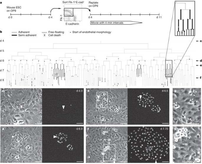
cell-tracking methods, we show that embryonic endothelial cells can be haemogenic. By continuous long-term single-cell observation of mouse mesodermal cells generating endothelial cell and
blood colonies, it was possible to detect haemogenic endothelial cells giving rise to blood cells. Living endothelial and haematopoietic cells were identified by simultaneous detection of
morphology and multiple molecular and functional markers. Detachment of nascent blood cells from endothelium is not directly linked to asymmetric cell division, and haemogenic endothelial
cells are specified from cells already expressing endothelial markers. These results improve our understanding of the developmental origin of mammalian blood and the potential generation of
haematopoietic stem cells from embryonic stem cells. Access through your institution Buy or subscribe This is a preview of subscription content, access via your institution ACCESS OPTIONS
Access through your institution Subscribe to this journal Receive 51 print issues and online access $199.00 per year only $3.90 per issue Learn more Buy this article * Purchase on
SpringerLink * Instant access to full article PDF Buy now Prices may be subject to local taxes which are calculated during checkout ADDITIONAL ACCESS OPTIONS: * Log in * Learn about
institutional subscriptions * Read our FAQs * Contact customer support SIMILAR CONTENT BEING VIEWED BY OTHERS GASTRULOIDS AS IN VITRO MODELS OF EMBRYONIC BLOOD DEVELOPMENT WITH SPATIAL AND
TEMPORAL RESOLUTION Article Open access 04 August 2022 EFFICIENT GENERATION OF HUMAN NOTCH LIGAND-EXPRESSING HAEMOGENIC ENDOTHELIAL CELLS AS INFRASTRUCTURE FOR IN VITRO HAEMATOPOIESIS AND
LYMPHOPOIESIS Article Open access 04 September 2024 CD32 CAPTURES COMMITTED HAEMOGENIC ENDOTHELIAL CELLS DURING HUMAN EMBRYONIC DEVELOPMENT Article Open access 09 April 2024 REFERENCES *
Godin, I. & Cumano, A. The hare and the tortoise: an embryonic haematopoietic race. _Nature Rev. Immunol._ 2, 593–604 (2002) Article CAS Google Scholar * Bertrand, J. Y. et al.
Characterization of purified intraembryonic hematopoietic stem cells as a tool to define their site of origin. _Proc. Natl Acad. Sci. USA_ 102, 134–139 (2005) Article ADS CAS Google
Scholar * Zovein, A. et al. Fate tracing reveals the endothelial origin of hematopoietic stem cells. _Cell Stem Cell_ 3, 625–636 (2008) Article CAS Google Scholar * Choi, K., Kennedy,
M., Kazarov, A., Papadimitriou, J. C. & Keller, G. A common precursor for hematopoietic and endothelial cells. _Development_ 125, 725–732 (1998) Article CAS Google Scholar * de
Bruijn, M. F. et al. Hematopoietic stem cells localize to the endothelial cell layer in the midgestation mouse aorta. _Immunity_ 16, 673–683 (2002) Article CAS Google Scholar * Jaffredo,
T., Gautier, R., Eichmann, A. & Dieterlen-Lievre, F. Intraaortic hemopoietic cells are derived from endothelial cells during ontogeny. _Development_ 125, 4575–4583 (1998) Article CAS
Google Scholar * Jaffredo, T. et al. From hemangioblast to hematopoietic stem cell: an endothelial connection? _Exp. Hematol._ 33, 1029–1040 (2005) Article Google Scholar * Nishikawa, S.
I. et al. _In vitro_ generation of lymphohematopoietic cells from endothelial cells purified from murine embryos. _Immunity_ 8, 761–769 (1998) Article CAS Google Scholar * Ueno, H. &
Weissman, I. L. Clonal analysis of mouse development reveals a polyclonal origin for yolk sac blood islands. _Dev. Cell_ 11, 519–533 (2006) Article CAS Google Scholar * Schroeder, T.
Imaging stem cell driven mammalian regeneration. _Nature_ 453, 345–351 (2008) Article ADS CAS Google Scholar * Fraser, S. T. et al. Definitive hematopoietic commitment within the
embryonic vascular endothelial-cadherin(+) population. _Exp. Hematol._ 30, 1070–1078 (2002) Article CAS Google Scholar * Sugiyama, D. et al. Erythropoiesis from acetyl LDL incorporating
endothelial cells at the preliver stage. _Blood_ 101, 4733–4738 (2003) Article CAS Google Scholar * Schroeder, T. et al. Recombination signal sequence-binding protein Jκ alters mesodermal
cell fate decisions by suppressing cardiomyogenesis. _Proc. Natl Acad. Sci. USA_ 100, 4018–4023 (2003) Article ADS CAS Google Scholar * Schroeder, T. et al. Activated Notch1 alters
differentiation of embryonic stem cells into mesodermal cell lineages at multiple stages of development. _Mech. Dev._ 123, 570–579 (2006) Article CAS Google Scholar * Hirashima, M.,
Kataoka, H., Nishikawa, S., Matsuyoshi, N. & Nishikawa, S. Maturation of embryonic stem cells into endothelial cells in an _in vitro_ model of vasculogenesis. _Blood_ 93, 1253–1263
(1999) Article CAS Google Scholar * Keller, G. Embryonic stem cell differentiation: emergence of a new era in biology and medicine. _Genes Dev._ 19, 1129–1155 (2005) Article CAS Google
Scholar * Nakano, T., Kodama, H. & Honjo, T. _In vitro_ development of primitive and definitive erythrocytes from different precursors. _Science_ 272, 722–724 (1996) Article ADS CAS
Google Scholar * Huber, T. L., Kouskoff, V., Fehling, H. J., Palis, J. & Keller, G. Haemangioblast commitment is initiated in the primitive streak of the mouse embryo. _Nature_ 432,
625–630 (2004) Article ADS CAS Google Scholar * North, T. E. et al. Prostaglandin E2 regulates vertebrate haematopoietic stem cell homeostasis. _Nature_ 447, 1007–1011 (2007) Article
ADS CAS Google Scholar * Kataoka, H. et al. Expressions of PDGF receptor alpha, c-Kit and Flk1 genes clustering in mouse chromosome 5 define distinct subsets of nascent mesodermal cells.
_Dev. Growth Differ._ 39, 729–740 (1997) Article CAS Google Scholar * Shalaby, F. et al. A requirement for Flk1 in primitive and definitive hematopoiesis and vasculogenesis. _Cell_ 89,
981–990 (1997) Article CAS Google Scholar * Yamashita, J. et al. Flk1-positive cells derived from embryonic stem cells serve as vascular progenitors. _Nature_ 408, 92–96 (2000) Article
ADS CAS Google Scholar * Guo, R., Sakamoto, H., Sugiura, S. & Ogawa, M. Endothelial cell motility is compatible with junctional integrity. _J. Cell. Physiol._ 211, 327–335 (2007)
Article CAS Google Scholar * Lampugnani, M. G. et al. A novel endothelial-specific membrane protein is a marker of cell–cell contacts. _J. Cell Biol._ 118, 1511–1522 (1992) Article CAS
Google Scholar * Voyta, J. C., Via, D. P., Butterfield, C. E. & Zetter, B. R. Identification and isolation of endothelial cells based on their increased uptake of acetylated-low density
lipoprotein. _J. Cell Biol._ 99, 2034–2040 (1984) Article CAS Google Scholar * Morita, K., Sasaki, H., Furuse, M. & Tsukita, S. Endothelial claudin: claudin-5/TMVCF constitutes tight
junction strands in endothelial cells. _J. Cell Biol._ 147, 185–194 (1999) Article CAS Google Scholar * Taoudi, S. et al. Extensive hematopoietic stem cell generation in the AGM region
via maturation of VE-cadherin+CD45+ pre-definitive HSCs. _Cell Stem Cell_ 3, 99–108 (2008) Article CAS Google Scholar * Ferkowicz, M. J. et al. CD41 expression defines the onset of
primitive and definitive hematopoiesis in the murine embryo. _Development_ 130, 4393–4403 (2003) Article CAS Google Scholar * Mikkola, H. K., Fujiwara, Y., Schlaeger, T. M., Traver, D.
& Orkin, S. H. Expression of CD41 marks the initiation of definitive hematopoiesis in the mouse embryo. _Blood_ 101, 508–516 (2003) Article CAS Google Scholar * Mitjavila-Garcia, M.
T. et al. Expression of CD41 on hematopoietic progenitors derived from embryonic hematopoietic cells. _Development_ 129, 2003–2013 (2002) Article CAS Google Scholar Download references
ACKNOWLEDGEMENTS We are grateful to S. Nishikawa, M. A. Rieger, A. Hermann and M. Yoder for technical advice and discussions, M. Ogawa and H. Niwa for the VEVC2 and EB3 ESC lines,
respectively, B. Schauberger for programming contributions, and A. Roth and C. Raithel for technical support. We thank M. Goetz, A. Hermann, M. A. Rieger and A. IJpenberg for critical
reading of the manuscript. Part of this study was financed by the Deutsche Forschungsgemeinschaft to T.S. and by the Leading Project for the Realization of Regenerative Medicine to S.-I.N.
AUTHOR CONTRIBUTIONS H.M.E. planned and performed experiments; S.-I.N. discussed results and commented on the manuscript; T.S. designed the study and experiments, developed the time-lapse
imaging and cell-tracking technology, performed initial experiments, analysed data with H.M.E. and wrote the paper with H.M.E. AUTHOR INFORMATION AUTHORS AND AFFILIATIONS * Institute of Stem
Cell Research, Helmholtz Center Munich—German Research Center for Environmental Health (GmbH), 85764 Neuherberg, Germany Hanna M. Eilken & Timm Schroeder * Laboratory for Stem Cell
Biology, RIKEN Center for Developmental Biology, 650-0047 Kobe, Japan Shin-Ichi Nishikawa Authors * Hanna M. Eilken View author publications You can also search for this author inPubMed
Google Scholar * Shin-Ichi Nishikawa View author publications You can also search for this author inPubMed Google Scholar * Timm Schroeder View author publications You can also search for
this author inPubMed Google Scholar CORRESPONDING AUTHOR Correspondence to Timm Schroeder. SUPPLEMENTARY INFORMATION SUPPLEMENTARY INFORMATION This file contains Supplementary Figures 1-8
with Legends, Supplementary Tables 1-3 and Supplementary Movies Legends 1-8 (PDF 2443 kb) SUPPLEMENTARY VIDEO 1 This time-lapse movie, covering a period of about 4.5 days, shows that cells
with endothelial morphology can give rise to blood cells (see file s1 for full legend). (MOV 9773 kb) SUPPLEMENTARY VIDEO 2 This time-lapse movie, covering a period of about 1 day, proofs
the cell identity of endothelial and blood cells by molecular and functional markers (see file s1 for full legend). (MOV 3788 kb) SUPPLEMENTARY VIDEO 3 This time-lapse movie, covering a
period of about 2.5 days, shows that nascent blood cells generated by endothelial cells express the surface marker CD41 prior to CD45 expression (see file s1 for full legend). (MOV 4070 kb)
SUPPLEMENTARY VIDEO 4 This time-lapse movie, covering a period of about 1.5 days, shows that mesodermal cells purified from 7.5 dpc mouse embryos can generate hemogenic endothelial cells
(see file s1 for full legend). (MOV 3892 kb) SUPPLEMENTARY VIDEO 5 This time-lapse movie, covering a period of about 6.5 days, shows that nascent blood cells generated by endothelial cells
have colony forming potential with high proliferative potential and multilineage (including megakaryocytic) potential. (MOV 8815 kb) SUPPLEMENTARY VIDEO 6 This time-lapse movie, covering a
period of about 5 hours, demonstrates the temporal and optical resolution of the primary image sequences used for cell tracking (see file s1 for full legend). (MOV 2331 kb) SUPPLEMENTARY
VIDEO 7 This time-lapse movie, covering a period of about 8.5 hours, demonstrates the temporal and optical resolution of the primary image sequences used for cell tracking (see file s1 for
full legend). (MOV 6633 kb) POWERPOINT SLIDES POWERPOINT SLIDE FOR FIG. 1 POWERPOINT SLIDE FOR FIG. 2 POWERPOINT SLIDE FOR FIG. 3 POWERPOINT SLIDE FOR FIG. 4 RIGHTS AND PERMISSIONS Reprints
and permissions ABOUT THIS ARTICLE CITE THIS ARTICLE Eilken, H., Nishikawa, SI. & Schroeder, T. Continuous single-cell imaging of blood generation from haemogenic endothelium. _Nature_
457, 896–900 (2009). https://doi.org/10.1038/nature07760 Download citation * Received: 29 May 2008 * Accepted: 09 January 2009 * Published: 01 February 2009 * Issue Date: 12 February 2009 *
DOI: https://doi.org/10.1038/nature07760 SHARE THIS ARTICLE Anyone you share the following link with will be able to read this content: Get shareable link Sorry, a shareable link is not
currently available for this article. Copy to clipboard Provided by the Springer Nature SharedIt content-sharing initiative

