Play all audios:
Excessive fructose intake has been associated with the development and progression of pancreatic cancer. This study aimed to elucidate the relationship between fructose utilization and
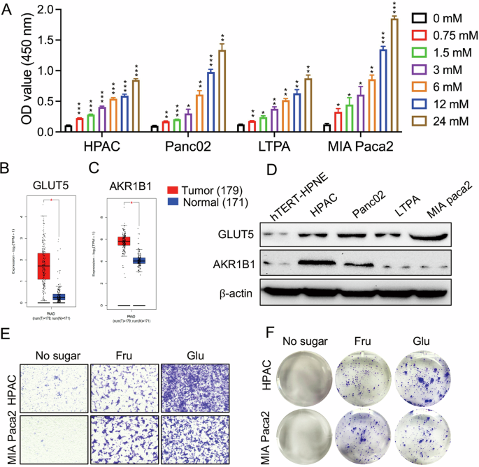
pancreatic cancer progression. Our findings revealed that pancreatic cancer cells have a high capacity to utilize fructose and are capable of converting glucose to fructose via the
AKR1B1-mediated polyol pathway, in addition to uptake via the fructose transporter GLUT5. Fructose metabolism exacerbates pancreatic cancer proliferation by enhancing glycolysis and
accelerating the production of key metabolites that regulate angiogenesis. However, pharmacological blockade of fructose metabolism has been shown to slow pancreatic cancer progression and
synergistically enhance anti-tumor capabilities when combined with anti-angiogenic agents. Overall, targeting fructose metabolism may prove to be a promising therapeutic approach in the
treatment of pancreatic cancer.
The datasets generated during and/or analyzed during the current study are available from the corresponding author on reasonable request.
This work was supported by Guangdong-Hong Kong-Macao Joint Laboratory for New Drug Screening (2023B1212120005).
Chinese Medicine Phenome Research Centre, School of Chinese Medicine, Hong Kong Baptist University, Hong Kong, China
Chengqiang Wang, Jiao Ma, Yitao Li, Xintong Yang, Aiping Lu, Kenneth C. P. Cheung & Wei Jia
Department of Pharmacology and Pharmacy, University of Hong Kong, Hong Kong, China
Center for Translational Medicine and Shanghai Key Laboratory of Diabetes Mellitus, Shanghai Sixth People’s Hospital Affiliated to Shanghai Jiao Tong University School of Medicine, Shanghai,
200233, China
NMPA Key Laboratory for Research and Evaluation of Drug Metabolism & Guangdong Provincial Key Laboratory of New Drug Screening & Guangdong-Hongkong-Macao Joint Laboratory for New Drug
Screening, School of Pharmaceutical Sciences, Southern Medical University, Guangzhou, 510515, China
Department of Experimental Medicine, University of Rome “Tor Vergata”, 00133, Rome, Italy
W.J. conceptualized the study. W.J and C.W. designed the study and coordinated the experimental planning. C.W. and L.W. performed the experiments. C.W., J.M., X.Y, and Y.L. were responsible
for mouse experiments. C.W. was responsible for cell studies. C.W. and L.W. were responsible for sample preparation and metabolomics analysis. C.W. K.C. and L.W. performed the data
preprocessing and statistical analysis. C.W. and W.J. drafted the manuscript and produced the figures. G.M., A.L., H.B., Q.Z., J.K. and K.C. provided valuable suggestions in data analysis
and interpretation. W.J. and C.W. critically revised the manuscript.
All animal experiments were approved by the Research Ethics Committee of the Hong Kong Baptist University (REC/20–21/0584) and were performed in accordance with relevant guidelines and
regulations. The relevant licenses were also approved by the Department of Health, Hong Kong, China (22-145 in DH/HT&A/8/2/6 Pt.6. and 21-189 in DH/HT&A/8/2/6 Pt.4.).
Publisher’s note Springer Nature remains neutral with regard to jurisdictional claims in published maps and institutional affiliations.
Springer Nature or its licensor (e.g. a society or other partner) holds exclusive rights to this article under a publishing agreement with the author(s) or other rightsholder(s); author
self-archiving of the accepted manuscript version of this article is solely governed by the terms of such publishing agreement and applicable law.
Anyone you share the following link with will be able to read this content: