Play all audios:
ABSTRACT Human noroviruses are the leading cause of severe childhood diarrhea worldwide, yet we know little about their pathogenic mechanisms. Murine noroviruses cause diarrhea in
interferon-deficient adult mice but these hosts also develop systemic pathology and lethality, reducing confidence in the translatability of findings to human norovirus disease. Herein we
report that a murine norovirus causes self-resolving diarrhea in the absence of systemic disease in wild-type neonatal mice, thus mirroring the key features of human norovirus disease and
representing a norovirus small animal disease model in wild-type mice. Intriguingly, lymphocytes are critical for controlling acute norovirus replication while simultaneously contributing to
disease severity, likely reflecting their dual role as targets of viral infection and key components of the host response. SIMILAR CONTENT BEING VIEWED BY OTHERS INFECTION OF NEONATAL MICE
WITH THE MURINE NOROVIRUS STRAIN WU23 IS A ROBUST MODEL TO STUDY NOROVIRUS PATHOGENESIS Article Open access 04 May 2023 AGE-ASSOCIATED FEATURES OF NOROVIRUS INFECTION ANALYSED IN MICE
Article 15 May 2023 ACUTE ROTAVIRUS INFECTION IS ASSOCIATED WITH THE INDUCTION OF CIRCULATING MEMORY CD4+ T CELL SUBSETS Article Open access 02 June 2023 INTRODUCTION Human noroviruses are
the leading cause of severe childhood diarrhea and gastroenteritis outbreaks worldwide1,2,3 yet we know very little about their pathogenic mechanisms. While human noroviruses cause modest
diarrhea in gnotobiotic piglets, gnotobiotic calves, and miniature piglets4,5,6, a limiting factor in studying norovirus pathogenesis is the lack of tractable small animal models that
recapitulate key features of disease observed in infected individuals. The first murine norovirus, MNV-1, was discovered nearly two decades ago7 and many other MNV strains have been reported
since then. MNV strains segregate into two phenotypic categories which differ in many aspects of pathogenesis, including their rate of clearance from infected hosts and cell tropism. MNV-1
is the prototype acute strain. It infects immune cells in the gut-associated lymphoid tissue (GALT) and reaches peak intestinal titers 1–2 days post-infection (dpi) with clearance from the
host by 7–14 dpi8. MNV-3 and MNV-CR6 are commonly referred to as persistent strains since they establish a life-long colonic infection9,10,11. The persistent reservoir is a rare type of
intestinal epithelial cell, the tuft cell12. While major strides have been made in understanding the cellular and tissue tropism of noroviruses using the MNV model system, a limitation of
the murine model is the lack of diarrhea in wild-type laboratory mice infected with any of the MNV strains studied to date13,14,15. While MNV-1 does not cause overt disease in adult
wild-type mice, infection of interferon (IFN)-deficient _Stat1__−/_− and _IFNαβγR__−/−_ adult mice causes severe weight loss and diarrhea similar to human norovirus-infected
individuals13,14,15,16. The persistent strains MNV-3 and MNV-CR6 cause less disease than MNV-1 in IFN-deficient mouse strains14,17. Like noroviruses, rotaviruses cause severe acute diarrhea
in people yet fail to cause disease in adult wild-type mice. However, it is well-established that neonatal wild-type mice are susceptible to rotavirus-induced diarrhea18,19,20. Moreover,
disease severity following human norovirus infection is greater in younger children21,22,23. Thus, we tested whether MNV causes disease in genetically wild-type neonatal mice. Herein, we
report that MNV-1 causes diarrhea in neonatal BALB/c mice while MNV-3 and MNV-CR6 cause attenuated disease in this model. In addition, we find that lymphocytes both contribute to disease
severity and are critical for controlling acute replication which likely reflects their dual role as both cellular targets and components of the host immune response. RESULTS DEVELOPMENT OF
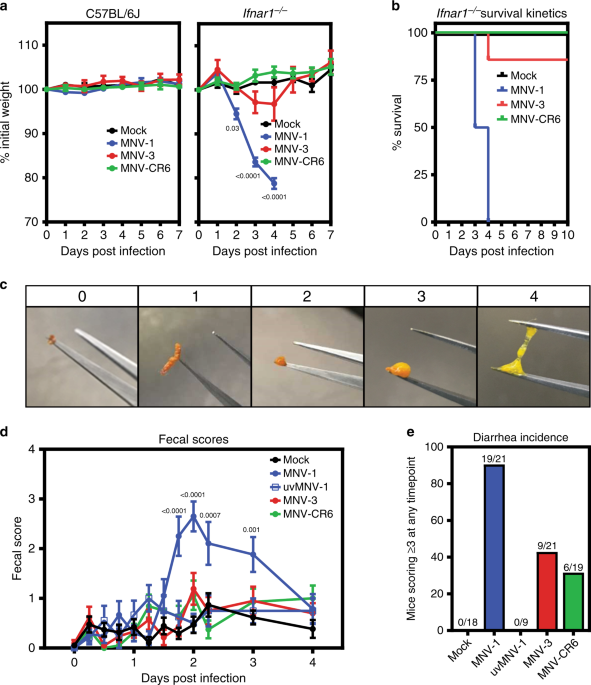
A NEONATAL MOUSE MODEL We first confirmed the established virulence profile of MNV strains in IFN-deficient adult mice by infecting adult wild-type or _Ifnar1__−/−_ mice with MNV-1, MNV-3,
or MNV-CR6. While MNV-1 caused weight loss and fatality of _Ifnar1__−/−_ mice by 4 dpi, MNV-3 and MNV-CR6 failed to cause overt disease (Fig. 1a, b). Overall, these results indicate that MNV
can cause disease in adult mice but virulence is regulated by an IFN response and viral genetic determinants. Moreover, MNV-1 infection of IFN-deficient mice causes systemic disease and
mortality in addition to diarrhea which are not hallmarks of human norovirus infection. Thus, we sought to develop a more relevant disease model by testing whether genetically wild-type
neonatal mice develop self-resolving diarrhea. Indeed, oral inoculation of 3-day-old BALB/c mice with acute MNV-1 caused diarrhea beginning at 42 h post-infection (hpi) and resolving by 4
dpi (Fig. 1c, d, and Supplementary Fig. 1c), with a 91% incidence (Fig. 1e). Virus replication was required for diarrhea induction, as demonstrated by the absence of disease in pups
inoculated with UV-inactivated MNV-1 (Fig. 1d, e and Supplementary Fig. 1b). Furthermore, MNV-1 diarrhea occurred in a dose-dependent manner (Fig. 2a, b). The incidence of diarrhea was
significantly higher in MNV-1-infected 3-day-old BALB/c mice compared with 4-day-old BALB/c mice, with a 91% versus 49% incidence, respectively (Fig. 2c, d), and slightly, although not
significantly, higher in female BALB/c mice than in male mice (Fig. 2e, f). MNV-1 induced diarrhea in BALB/c mice at two independent research institutions (University of Florida and the
National Institutes of Health). For all other experiments described herein, we used 3-day-old BALB/c mice of mixed sex. We next tested whether the persistent strains MNV-3 and MNV-CR6
likewise caused diarrhea in neonatal mice. Compared with the 91% incidence of diarrhea in MNV-1-infected BALB/c pups, MNV-3 and MNV-CR6 caused diarrhea in 43% and 32% of pups, respectively
(Fig. 1d, e and Supplementary Fig. 1d, e). Collectively, these data reveal that MNV-1 infection causes self-resolving acute diarrhea in neonatal BALB/c mice and that MNV-3 and MNV-CR6 cause
a reduced incidence of diarrhea relative to MNV-1, mirroring their virulence patterns previously observed in adult IFN-deficient mice14,17. MNV INFECTION INDUCES PATHOLOGICAL CHANGES IN
NEONATAL MICE We next determined whether MNV-induced diarrhea is associated with histopathological changes along the intestinal tract. A number of human volunteer studies have been carried
out where intestinal biopsies were obtained from subjects at the time of norovirus symptoms24,25,26,27,28,29. While the epithelium remained intact with no gross lesions and there was only a
minimal to moderate increase in lamina propria cellularity noted in these patients, there were consistent infection-associated pathologies including villous blunting and broadening,
vacuolated and disorganized epithelial cells, and crypt cell hyperplasia. Histopathological changes in MNV-infected neonatal mice are entirely consistent with these human biopsies: While the
epithelium itself remained intact and only modest inflammation was observed, there was villous broadening, epithelial disorganization and vacuolization, and crypt cell hyperplasia (Fig. 3a
and Supplementary Fig. 2). We also observed lacteal dilation, a pathology previously noted in MNV-4-infected _Stat1__−/−_ mice30 that can be indicative of impaired fat absorption. Overall
pathology was more pronounced in the proximal half of the small intestine than the distal half, and it positively correlated with diarrhea incidence when comparing intestinal sections from
pups infected with MNV-1, MNV-3, and MNV-CR6 (Fig. 3b, c and Supplementary Fig. 2). MNV-1 REPLICATES IN SUBEPITHELIAL CELLS OF THE SMALL INTESTINE Because MNV-1 caused the highest incidence
of diarrhea in neonates, we further characterized its pathogenesis in this model. Virus titers were similar in all regions of the gastrointestinal tract at the peak of disease, 2 dpi, and
splenic titers were high at this time point (Fig. 4a). In order to confirm viral replication in vivo, we infected neonatal mice with light-sensitive neutral red-labeled MNV-1 which enables
differentiation between input and newly synthesized virus31,32. Newly synthesized virus was detectable in all tissues as early as 0.5 dpi and peaked at 1 dpi (Fig. 4b). We next examined the
cell tropism of MNV-1 during symptomatic infection. We previously showed that MNV-1 infects immune cells in the GALT of the distal small intestine of wild-type adult mice8. To determine
whether there is a similar tropism during symptomatic norovirus infection, intestinal sections from MNV-1-infected neonates were hybridized with probes to the viral positive- and
negative-sense RNA species and analyzed by RNAscope-based in situ hybridization. At 1 dpi, the peak of viral titers, viral positive-sense RNA was detected in both epithelial and
subepithelial cells in the intestinal villi and GALT but negative-sense RNA indicative of viral replication was exclusively detected in subepithelial cells in the GALT and the lamina propria
(Fig. 5a, b). The presence of viral positive-sense, but not negative-sense, viral RNA in intestinal epithelial cells suggests that either virions can enter these cells but not replicate; or
that replication occurs in these cells at a level below the limit of detection of our assay. Substantial viral replication was detected in splenocytes, consistent with high splenic titers
and with a predominantly immune cell tropism. At 2 dpi, the peak of diarrhea, there was still viral positive-sense RNA along the gastrointestinal tract but minimal negative-sense viral RNA,
demonstrating that viral replication in the gut has primarily been controlled by this time point (Fig. 5c–e). The pattern of viral positive-sense RNA at this time point was variable among
the seven pups analyzed: Four pups contained positive-sense RNA primarily in subepithelial cells of the GALT and lamina propria similar to 1 dpi (Fig. 5c) whereas two pups contained
positive-sense viral RNA primarily in villous epithelial cells (Fig. 5d). Intriguingly, this signal was confined to the distal region of the small intestine and to the upper half of the
villi in this region. The remaining pup displayed an intermediate phenotype, with positive-sense RNA detected in subepithelial and epithelial cells. All pups had substantial positive-sense
RNA and variable amounts of negative-sense RNA in the spleen at 2 dpi. Overall, viral replication was observed in subepithelial cells in the intestine and spleen. LYMPHOCYTES EXHIBIT DUAL
ROLES DURING ACUTE MNV-1 INFECTION To begin dissecting the role of specific cellular targets in norovirus disease, we infected lymphocyte-deficient _Rag1__−/−_ neonates with MNV-1.
Surprisingly, virus titers at 2 dpi were significantly higher in _Rag1__−/−_ mice compared with wild-type controls (Fig. 6a). In spite of these increased virus titers, the incidence of
diarrhea was reduced in _Rag1__−/−_ mice (56%) compared with wild-type mice (90%) (Fig. 6b, c). These collective results suggest that lymphocytes play opposing roles during acute norovirus
infection, being important for control of MNV-1 replication while also contributing to disease severity possibly related to their permissiveness to the virus. DISCUSSION The establishment of
a small animal model to study norovirus-induced diarrhea in a genetically wild-type host that recapitulates key features of human disease will revolutionize the field’s ability to
understand the precise mechanisms underlying norovirus virulence and to rationally design antiviral therapies. Strengths of this neonatal mouse model compared with adult IFN-deficient mice
are that disease is limited to the intestinal tract and self-resolving. Disease severity in this model is (i) regulated by viral genetics, (ii) age-dependent, (iii) not associated with
disruption of the intestinal epithelium or notable inflammation, and (iv) associated with immune cell infection. In particular, the finding that highly genetically related MNV strains
display differences in diarrhea severity will facilitate identification of viral virulence determinants. Underscoring the potential of this neonatal mouse model system, the discovery that
neonatal mice are susceptible to rotavirus diarrhea was key to defining viral disease mechanisms such as the roles of rotavirus NSP4 as a viral enterotoxin33 and rotavirus stimulation of the
enteric nervous system leading to increased intestinal transit associated with diarrhea34,35. The model system described herein holds a similar promise to reveal additional insights into
norovirus disease mechanisms. METHODS CELLS AND VIRUSES Cell lines used in this study tested negative for mycoplasma contamination and were not authenticated. The RAW 264.7 cell line (ATCC)
was maintained in Dulbecco’s modified Eagle medium (DMEM; Fisher Scientific) supplemented with 10% fetal bovine serum (FBS, Atlanta Biologicals), 100 U penicillin/mL, and 100 μg/mL
streptomycin. Stocks of recombinant MNV-1.CW3 (GenBank accession number KC782764, referred to as MNV-1), MNV-3 (GenBank accession number KC792553), and MNV-CR6 (GenBank accession number
JQ237823.1) were generated36. 293T cells were seeded at 106 cells per well in a 6-well culture plate. The next day, 106 293T cells were transfected with 5 µg infectious clone using
Lipofectamine 2000 (Life Technologies), cells were lysed by freeze-thaw at −80 °C after 1 day, and lysates titrated with a standard TCID50 assay11 as described below and applied to RAW 264.7
cells. RAW 264.7 lysates were freeze-thawed when cultures displayed 90% cytopathic effect and supernatants were clarified by low-speed centrifugation followed by purification through a 25%
sucrose cushion. The viral genomes of stocks were sequenced completely to confirm no mutations arose during stock generation and titered by a standard TCID50 assay11. For the TCID50 assay,
eight replicates each of multiple dilutions per sample were applied to RAW 264.7 cells and cytopathicity scored at 7 dpi. Three independent experiments were averaged to determine stock
titers. A mock inoculum stock was prepared using RAW 264.7 lysate from uninfected cultures. A neutral red-labeled MNV-1 stock was generated by infecting RAW 264.7 cells at a MOI 0.05 with
MNV-1 in the presence of 10 μg/ml neutral red (Sigma), a light-sensitive dye31,37. After 2 days, supernatant was collected in a darkened room using a red photolight (Premier OMNI). Stocks
were freeze-thawed twice and stored in a light safe box at −80 °C. Viral titer was determined by neutral red virus plaque assay without white light exposure. For UV inactivation of MNV-1,
MNV-1 was exposed to 250,000 µJ cm−2 UV for 30 min. UV inactivation was confirmed using TCID50 assay. MICE Specific-pathogen-free (SPF) mice used in this study were bred and housed in animal
facilities at the University of Florida, National Institutes of Health, and University of Michigan. All animal experiments were performed in strict accordance with federal and university
guidelines. The animal protocols were approved by the Institutional Animal Care and Use Committee at the University of Florida (study number 20190632), National Institutes of Health (study
number H-0299), and University of Michigan (study number 00006658). The conditions in animal rooms used in this study fall within the standards set by the “Guide for the Care and Use of
Laboratory Animals”. Specifically, the average temperature is 72 °F and always is within 68–79 °F. Humidity ranges from 30 to 70%. The room light cycle is 12-h on and 12-h off. Experiments
were not performed in a blinded fashion nor was randomization used. Sample sizes were determined using power calculations based on our prior experience with the MNV system. Age- and
sex-matched adult C57BL/6J (Jackson no. 000664) and C57BL/6J-_Ifnar1__−/−_ (Jackson no. 010830) mice were used in adult mouse infections. Litters of 3- or 4-day-old BALB/c (Charles River no.
028) and 3-day-old BALB/c-_Rag1__−/−_ (Jackson no. 003145) pups including males and females were used in neonatal mouse infections. At least two independent litters were infected per
condition in each experiment. MOUSE INFECTIONS AND OTHER IN VIVO PROCEDURES For all adult MNV infection experiments, 6- to 12-week-old, sex-matched mice were inoculated perorally (p.o.) with
107 TCID50 units of the indicated virus strain. For virulence assays in adult mice, C57BL/6J and C57BL/6J-_Ifnar1__−/−_ mice were observed for weight loss and survival daily for up to 14
days. The daily weights were compared with day 0 weights to calculate the relative weight loss. For all neonatal mouse experiments, BALB/c or BALB/c-_Rag1__−/−_ mice were infected at 3 or 4
days of age with the indicated dose of the indicated virus strain in a volume of 40 μl delivered by oral gavage using a 22ga plastic feeding tube. Mice were observed every 6 h for the first
54 h post-infection (hpi), then daily thereafter until 4 dpi. Approximately 25% of pups failed to survive to the end of the experiment. A comparable frequency of lethality was observed in
mock-inoculated and MNV-infected groups (see Source data file) so we presume that it resulted from stress associated with handling of the pups during the inoculation procedure either on the
pup itself or the dam. This is not unexpected when handling pups as young as 3 days of age. At each time point, the abdomen of each mouse was palpated to induce defecation. Fecal condition
was assessed based on color and consistency according to a 5-point scale: 0, no defecation or solid; 1, firm, orange, does not smear; 2, pasty, orange or mixed color, does not smear; 3,
orange or yellow, semi-liquid and smears; 4, yellow, liquid, and smears. For the purpose of calculating the incidence of diarrhea, any pup that received a score of 3 or 4 at any time point
was considered positive for disease. For MNV titer determination, tissue samples were harvested and titrated by plaque assay14,36. For the plaque assays, tissues were weighed and homogenized
by bead beating using 1.0 mm zirconia/silica beads. Dilutions of tissue samples were prepared and applied to RAW 264.7 cells. Plates were incubated for 1 h at room temperature while rocking
followed by an overlay of 1.5% SeaPlaque Agarose and modified eagle medium (MEM) supplemented with 10% fetal bovine serum (FBS, Atlanta Biologicals), 100 U penicillin/mL, and 100 μg/mL
streptomycin. After 2 days for MNV-1 and 3 days for MNV-3 and MNV-CR6, cells were stained with 1.5% SeaKem Agarose in phosphate-buffered saline (PBS) to visualize plaques. Plaques were
counted 3–4 h later. In certain experiments, neutral red-labeled virus was used in order to differentiate between inoculum and newly synthesized virus along the intestinal tract31,37. When
using tissues from mice infected with neutral red virus, samples were exposed to white light for 30 min prior to titration in order to inactivate input virus. HISTOLOGY Small and large
intestinal tissue sections as well as spleen sections were collected from 3-day-old pups infected with mock inoculum or 108 TCID50 units MNV-1, MNV-3, or MNV-CR6 at 1 and 2 dpi. Intestinal
issues were swiss-rolled and fixed in 10% buffered formalin for 16 h, transferred to PBS, and then paraffin-embedded and sectioned by the University of Florida Molecular Pathology Core.
Serial sections of 4-µm thickness were cut from each tissue and slides stained with hematoxylin and eosin by the University of Florida Molecular Pathology Core. Tissue sections were imaged
using a Nikon Ti-E widefield microscope with a Nikon DS-Fi2 color camera and NIS Elements software at the University of Florida Cell and Tissue Analysis Core. Histology slides were scored
blindly by an animal veterinarian based on an established scoring system38. Three categories—inflammatory cell infiltrate, epithelial changes, and mucosal architecture—were scored for two
criteria each: Inflammatory cell infiltrate was scored for severity (0 = none; 1 = minimal; 2 = mild with scattered polymorphonuclear neutrophils; 3 = moderate) and extent (0 = none; 1 =
mucosal; 2 = mucosal and submucosal). Epithelial changes were scored for crypt hyperplasia (0 = 2–3 mm crypt height at 10X; 1 = 3–4 mm crypt height; 2 = 4–5 mm crypt height; and 3 = ≥5 mm
crypt height) and reduction in goblet cells (0 = no reduction; 1 = minimal reduction). Mucosal architecture was scored for vacuolated epithelium (0 = none; 1 = present at villous tip; 2 =
present and disorganized, seen at the base) and villous tip swelling (0 = none; 1 = lacteal separation with cellular material in the center; 2 = lacteal dilation). Data are presented as the
total pathology score for all criteria per mouse averaged per group (Fig. 3b, c) and separated by category (Supplementary Fig. 2). RNASCOPE ISH RNAscope ISH assays were performed using the
RNAscope 2.5 HD Assay-RED kit according to manufacturer’s instructions (Advanced Cell Diagnostics, Newark, CA). Formalin-fixed, paraffin-embedded serial sections of intestinal swiss rolls
from mock- and MNV-1-infected pups were deparaffinized by heating at 60 °C for 30 min followed by xylene treatment and dehydration. Sections were hybridized with custom-designed probes
targeting positive-sense or negative-sense MNV-1 RNA for 2 h at 40 °C prior to probe amplification and detection8. Sections were counterstained with 50% hematoxylin to visualize tissue
morphology and imaged using a Nikon DS-Fi2 color camera and NIS Elements software at the University of Florida Cell and Tissue Analysis Core. Sections were scored based on amount of virus
present in the GALT, intestinal lamina propria, and intestinal epithelial cells on a scale from 0 to 3 (0 = 0–1 dots, 1 = rare staining but at least 2 dots, 2 = consistent, distinguishable
dots throughout the section, 3 = intense staining with overlapping dots). Positive (PPIB) and negative (DapB) control probes were stained in parallel for all experiments, and six mock mice
were stained. STATISTICAL ANALYSES All data were analyzed with GraphPad Prism software. Error bars denote standard errors of mean in all figures and _P_ values were determined using one- or
two-way ANOVA with corrections for multiple comparisons. In the case of survival curves and incidence of diarrhea, statistical significance was determined using a Mantel-Cox test. REPORTING
SUMMARY Further information on research design is available in the Nature Research Reporting Summary linked to this article. DATA AVAILABILITY All data that support the findings of this
study are included in this paper, the Supplementary information, the Source data file, and/or available from the corresponding author on request. The source data underlying Fig. 1a, b, d, e,
Fig. 2a–f, Fig. 3b, c, Fig. 4a, b, Fig. 5b, e, Fig. 6a–c, Supplementary Fig. 1a–e, and Supplementary Fig. 2a–c are provided as a source data file. REFERENCES * Koo, H. L. et al.
Noroviruses: the most common pediatric viral enteric pathogen at a large university hospital after introduction of rotavirus vaccination. _J. Pediatr. Infect. Dis. Soc._ 2, 57–60 (2013).
Article Google Scholar * Patel, M. M. Systematic literature review of role of noroviruses in sporadic gastroenteritis. _Emerg. Infect. Dis._ 14, 1224–1231 (2008). Article Google Scholar
* Ahmed, S. M. et al. Global prevalence of norovirus in cases of gastroenteritis: a systematic review and meta-analysis. _Lancet Infect. Dis._ 14, 725–730 (2014). Article Google Scholar *
Cheetham, S. et al. Pathogenesis of a genogroup II human norovirus in gnotobiotic pigs. _J. Virol._ 80, 10372–10381 (2006). Article CAS Google Scholar * Seo, D. J. et al. Experimental
miniature piglet model for the infection of human norovirus GII. _J. Med. Virol._ 90, 655–662 (2018). Article CAS Google Scholar * Souza, M., Azevedo, M. S. P., Jung, K., Cheetham, S.
& Saif, L. J. Pathogenesis and immune responses in gnotobiotic calves after infection with the genogroup II.4-HS66 strain of human norovirus. _J. Virol._ 82, 1777–1786 (2008). Article
CAS Google Scholar * Karst, S. M., Wobus, C. E., Lay, M., Davidson, J. & Virgin, H. W. STAT1-dependent innate immunity to a norwalk-like virus. _Science_ 299, 1575–1578 (2003). Article
ADS CAS Google Scholar * Grau, K. R. et al. The major targets of acute norovirus infection are immune cells in the gut-associated lymphoid tissue. _Nat. Microbiol._ 2, 1586 (2017).
Article CAS Google Scholar * Hsu, C. C., Riley, L. K., Wills, H. M. & Livingston, R. S. Persistent infection with and serologic crossreactivity of three novel murine noroviruses.
_Comp. Med._ 56, 247–251 (2006). CAS PubMed Google Scholar * Arias, A., Bailey, D., Chaudhry, Y. & Goodfellow, I. G. Development of a reverse genetics system for murine norovirus 3;
long-term persistence occurs in the caecum and colon. _J. Gen. Virol._ 93, 1432–1441 (2012). Article CAS Google Scholar * Thackray, L. B. et al. Murine noroviruses comprising a single
genogroup exhibit biological diversity despite limited sequence divergence. _J. Virol._ 81, 10460–10473 (2007). Article CAS Google Scholar * Wilen, C. B. et al. Tropism for tuft cells
determines immune promotion of norovirus pathogenesis. _Science_ 360, 204–208 (2018). Article ADS CAS Google Scholar * Mumphrey, S. M. et al. Murine norovirus 1 infection is associated
with histopathological changes in immunocompetent hosts, but clinical disease is prevented by STAT1-dependent interferon responses. _J. Virol._ 81, 3251–3263 (2007). Article CAS Google
Scholar * Kahan, S. M. et al. Comparative murine norovirus studies reveal a lack of correlation between intestinal virus titers and enteric pathology. _Virology_ 421, 202–210 (2011).
Article CAS Google Scholar * Zhu, S. et al. Regulation of norovirus virulence by the VP1 protruding domain correlates with B cell infection efficiency. _J. Virol._ 90, 2858–2867 (2016).
Article CAS Google Scholar * Rocha-Pereira, J., Kolawole, A. O., Verbeken, E., Wobus, C. E. & Neyts, J. Post-exposure antiviral treatment of norovirus infections effectively protects
against diarrhea and reduces virus shedding in the stool in a mortality mouse model. _Antivir. Res._ 132, 76–84 (2016). Article CAS Google Scholar * Strong, D. W., Thackray, L. B., Smith,
T. J. & Virgin, H. W. Protruding domain of capsid protein is necessary and sufficient to determine murine norovirus replication and pathogenesis in vivo. _J. Virol._ 86, 2950–2958
(2012). Article CAS Google Scholar * Feng, N., Franco, M. A. & Greenberg, H. B. Murine model of rotavirus infection. _Adv. Exp. Med. Biol._ 412, 233–240 (1997). Article CAS Google
Scholar * Ramig, R. F. The effects of host age, virus dose, and virus strain on heterologous rotavirus infection of suckling mice. _Microb. Pathog._ 4, 189–202 (1988). Article CAS Google
Scholar * Du, J. et al. Detailed analysis of BALB/c mice challenged with wild type rotavirus EDIM provide an alternative for infection model of rotavirus. _Virus Res._ 228, 134–140 (2017).
Article CAS Google Scholar * Cannon, J. L., Lopman, B. A., Payne, D. C. & Vinjé, J. Birth cohort studies assessing norovirus infection and immunity in young children: a review. _Clin.
Infect. Dis._ 69, 357–365 (2019). Article CAS Google Scholar * Menon, V. K. et al. Norovirus gastroenteritis in a birth cohort in southern India. _PLoS ONE_ 11, e0157007 (2016). Article
Google Scholar * Rouhani, S. et al. Norovirus infection and acquired immunity in 8 countries: results from the MAL-ED study. _Clin. Infect. Dis._ 62, 1210–1217 (2016). Article Google
Scholar * Agus, S. G., Dolin, R., Wyatt, R. G., Tousimis, A. J. & Northrup, R. S. Acute infectious nonbacterial gastroenteritis: intestinal histopathology. Histologic and enzymatic
alterations during illness produced by the Norwalk agent in man. _Ann. Intern. Med._ 79, 18–25 (1973). Article CAS Google Scholar * Schreiber, D. S., Blacklow, N. R. & Trier, J. S.
The small intestinal lesion induced by Hawaii agent acute infectious nonbacterial gastroenteritis. _J. Infect. Dis._ 129, 705–708 (1974). Article CAS Google Scholar * Parrino, T. A.,
Schreiber, D. S., Trier, J. S., Kapikian, A. Z. & Blacklow, N. R. Clinical immunity in acute gastroenteritis caused by Norwalk agent. _N. Engl. J. Med._ 297, 86–89 (1977). Article CAS
Google Scholar * Schreiber, D. S., Blacklow, N. R. & Trier, J. S. The mucosal lesion of the proximal small intestine in acute infectious nonbacterial gastroenteritis. _N. Engl. J. Med._
288, 1318–1323 (1973). Article CAS Google Scholar * Dolin, R., Levy, A. G., Wyatt, R. G., Thornhill, T. S. & Gardner, J. D. Viral gastroenteritis induced by the Hawaii agent. Jejunal
histopathology and serologic response. _Am. J. Med._ 59, 761–768 (1975). Article CAS Google Scholar * Troeger, H. et al. Structural and functional changes of the duodenum in human
norovirus infection. _Gut_ 58, 1070–1077 (2009). Article CAS Google Scholar * Seamons, A. et al. Obstructive lymphangitis precedes colitis in murine norovirus–infected Stat1-deficient
mice. _Am. J. Pathol._ 188, 1536–1554 (2018). Article Google Scholar * González-Hernández, M. B., Perry, J. W. & Wobus, C. E. Neutral red assay for murine norovirus replication and
detection in a mouse. _Bio-Protoc_. 3, e415 (2013). * Perry, J. W. & Wobus, C. E. Endocytosis of murine norovirus 1 into murine macrophages is dependent on dynamin II and cholesterol.
_J. Virol._ 84, 6163–6176 (2010). Article CAS Google Scholar * Ball, J. M., Tian, P., Zeng, C. Q., Morris, A. P. & Estes, M. K. Age-dependent diarrhea induced by a rotaviral
nonstructural glycoprotein. _Science_ 272, 101–104 (1996). Article ADS CAS Google Scholar * Lundgren, O. et al. Role of the enteric nervous system in the fluid and electrolyte secretion
of rotavirus diarrhea. _Science_ 287, 491–495 (2000). Article ADS CAS Google Scholar * Istrate, C., Hagbom, M., Vikström, E., Magnusson, K.-E. & Svensson, L. Rotavirus infection
increases intestinal motility but not permeability at the onset of diarrhea. _J. Virol._ 88, 3161–3169 (2014). Article Google Scholar * Zhu, S. et al. Identification of immune and viral
correlates of norovirus protective immunity through comparative study of intra-cluster norovirus strains. _PLoS Pathog._ 9, e1003592 (2013). Article CAS Google Scholar *
Gonzalez-Hernandez, M. B. et al. Efficient norovirus and reovirus replication in the mouse intestine requires microfold (M) cells. _J. Virol._ 88, 6934–6943 (2014). Article Google Scholar
* Erben, U. et al. A guide to histomorphological evaluation of intestinal inflammation in mouse models. _Int. J. Clin. Exp. Pathol._ 7, 4557–4576 (2014). PubMed PubMed Central Google
Scholar Download references ACKNOWLEDGEMENTS S.M.K. was funded by NIH R01AI116892, NIH R01AI081921, and NIH R01AI141478. A.N.R. was supported by NIH 5T32AI007110. E.W.H. was supported by
NIH T90DE021990. C.E.W. was funded by NIH R01AI141478 and the Biological Sciences Scholars Program at the University of Michigan. AUTHOR INFORMATION Author notes * These authors contributed
equally: Alexa N. Roth, Emily W. Helm. AUTHORS AND AFFILIATIONS * Department of Molecular Genetics & Microbiology, College of Medicine, University of Florida, Gainesville, FL, USA Alexa
N. Roth, Emily W. Helm, Erin Kirsche, Jonathan C. Smith & Stephanie M. Karst * Department of Microbiology and Immunology, University of Michigan, Ann Arbor, MI, USA Carmen Mirabelli
& Christiane E. Wobus * Office of Research, College of Medicine, University of Florida, Gainesville, FL, USA Laura B. Eurell * Laboratory of Host-Pathogen Dynamics, National Heart Lung
and Blood Institute, National Institutes of Health, Bethesda, MD, USA Sourish Ghosh & Nihal Altan-Bonnet Authors * Alexa N. Roth View author publications You can also search for this
author inPubMed Google Scholar * Emily W. Helm View author publications You can also search for this author inPubMed Google Scholar * Carmen Mirabelli View author publications You can also
search for this author inPubMed Google Scholar * Erin Kirsche View author publications You can also search for this author inPubMed Google Scholar * Jonathan C. Smith View author
publications You can also search for this author inPubMed Google Scholar * Laura B. Eurell View author publications You can also search for this author inPubMed Google Scholar * Sourish
Ghosh View author publications You can also search for this author inPubMed Google Scholar * Nihal Altan-Bonnet View author publications You can also search for this author inPubMed Google
Scholar * Christiane E. Wobus View author publications You can also search for this author inPubMed Google Scholar * Stephanie M. Karst View author publications You can also search for this
author inPubMed Google Scholar CONTRIBUTIONS A.N.R., E.W.H., and S.M.K. designed the study and analyzed results. A.N.R., E.W.H., S.G., and C.M. performed mouse infections, harvests, and
plaque assays. E.K. and J.C.S. performed plaque assays. L.B.E. scored histology slides in a blinded fashion. A.N.R., E.W.H., and S.M.K. prepared the paper. C.E.W. and N.A-B. edited the
paper. CORRESPONDING AUTHOR Correspondence to Stephanie M. Karst. ETHICS DECLARATIONS COMPETING INTERESTS The authors declare no competing interests. ADDITIONAL INFORMATION PEER REVIEW
INFORMATION _Nature Communications_ thanks the anonymous reviewers for their contribution to the peer review of this work. PUBLISHER’S NOTE Springer Nature remains neutral with regard to
jurisdictional claims in published maps and institutional affiliations. SUPPLEMENTARY INFORMATION SUPPLEMENTARY INFORMATION REPORTING SUMMARY SOURCE DATA SOURCE DATA RIGHTS AND PERMISSIONS
OPEN ACCESS This article is licensed under a Creative Commons Attribution 4.0 International License, which permits use, sharing, adaptation, distribution and reproduction in any medium or
format, as long as you give appropriate credit to the original author(s) and the source, provide a link to the Creative Commons license, and indicate if changes were made. The images or
other third party material in this article are included in the article’s Creative Commons license, unless indicated otherwise in a credit line to the material. If material is not included in
the article’s Creative Commons license and your intended use is not permitted by statutory regulation or exceeds the permitted use, you will need to obtain permission directly from the
copyright holder. To view a copy of this license, visit http://creativecommons.org/licenses/by/4.0/. Reprints and permissions ABOUT THIS ARTICLE CITE THIS ARTICLE Roth, A.N., Helm, E.W.,
Mirabelli, C. _et al._ Norovirus infection causes acute self-resolving diarrhea in wild-type neonatal mice. _Nat Commun_ 11, 2968 (2020). https://doi.org/10.1038/s41467-020-16798-1 Download
citation * Received: 24 October 2019 * Accepted: 25 May 2020 * Published: 11 June 2020 * DOI: https://doi.org/10.1038/s41467-020-16798-1 SHARE THIS ARTICLE Anyone you share the following
link with will be able to read this content: Get shareable link Sorry, a shareable link is not currently available for this article. Copy to clipboard Provided by the Springer Nature
SharedIt content-sharing initiative