Play all audios:
Double-β-decay involves the simultaneous conversion of two neutrons into two protons, and the emission of two electrons and two neutrinos; the neutrinoless process, although not yet
observed, is thought to involve the emission of the two electrons but no neutrinos. The search for neutrinoless-double-β-decay probes fundamental properties of neutrinos, including whether
or not the neutrino and antineutrino are distinct particles. Double-β-decay detectors are large and expensive, so it is essential to achieve the highest possible sensitivity with each study,
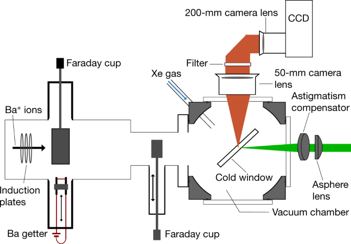
and removing spurious contributions (‘background’) from detected signals is crucial. In the nEXO neutrinoless-double-β-decay experiment, the identification, or ‘tagging’, of the 136Ba
daughter atom resulting from the double-β decay of 136Xe provides a technique for discriminating background. The tagging scheme studied here uses a cryogenic probe to trap the barium atom in
a solid xenon matrix, where the barium atom is tagged through fluorescence imaging. Here we demonstrate the imaging and counting of individual barium atoms in solid xenon by scanning a
focused laser across a solid xenon matrix deposited on a sapphire window. When the laser irradiates an individual atom, the fluorescence persists for about 30 seconds before dropping
abruptly to the background level—a clear confirmation of one-atom imaging. Following evaporation of a barium deposit, the residual barium fluorescence is 0.16 per cent or less. Our technique
achieves the imaging of single atoms in a solid noble element, establishing the basic principle of barium tagging for nEXO.
Source Data for Figs. 2–6 are provided with the online version of the paper.
We thank Picoquant for the loan of time-resolved-photon-counting equipment. Discussions with J. G. McCaffrey, B. Gervais and A. van Orden are appreciated. This material is based upon work
supported by the National Science Foundation under grant number PHY-1649324 and the US Department of Energy, Office of Science, Office of High Energy Physics under award number
DE-FG02-03ER41255.
Nature thanks Mark Chen, John McCaffrey and the other anonymous reviewer(s) for their contribution to the peer review of this work.
A list of participants and their affiliations appears at the end of the paper.
Physics Department, Colorado State University, Fort Collins, CO, USA
C. Chambers, T. Walton, D. Fairbank, A. Craycraft, D. R. Yahne, J. Todd, A. Iverson & W. Fairbank
Department of Physics, Carleton University, Ottawa, Ontario, Canada
A. Alamre, I. Badhrees, W. Cree, R. Gornea, C. Jessiman, T. Koffas, B. Veenstra & J. Watkins
Department of Physics and Center for Exploration of Energy and Matter (CEEM), Indiana University, Bloomington, IN, USA
Erlangen Centre for Astroparticle Physics (ECAP), Friedrich-Alexander University Erlangen-Nürnberg, Erlangen, Germany
G. Anton, J. Hößl, P. Hufschmidt, T. Michel, S. Schmidt, J. Schneider, M. Wagenpfeil, G. Wrede & T. Ziegler
I. J. Arnquist, E. W. Hoppe, J. L. Orrell, G. S. Ortega, C. T. Overman, R. Saldanha & R. Tsang
King Abdulaziz City for Science and Technology (KACST), Riyadh, Saudi Arabia
Department of Physics, Duke University, and Triangle Universities Nuclear Laboratory (TUNL), Durham, NC, USA
Physics Department, University of Illinois, Urbana-Champaign, IL, USA
Institute for Theoretical and Experimental Physics named by A. I. Alikhanov of National Research Center ‘Kurchatov Institute’, Moscow, Russia
V. Belov, A. Burenkov, A. Karelin, A. Kuchenkov, V. Stekhanov & O. Zeldovich
Department of Physics, University of South Dakota, Vermillion, SD, USA
F. Bourque, S. A. Charlebois, M. Côté, R. Fontaine, F. Nolet, S. Parent, J.-F. Pratte, T. Rossignol, N. Roy, G. St-Hilaire & F. Vachon
J. P. Brodsky, M. Heffner, A. House, S. Sangiorgio & T. Stiegler
Department of Physics, Applied Physics and Astronomy, Rensselaer Polytechnic Institute, Troy, NY, USA
Physics Department, McGill University, Montreal, Québec, Canada
T. Brunner, J. Dilling, G. Gallina, R. Gornea, R. Krücken, Y. Lan & F. Retière
G. F. Cao, W. R. Cen, Y. Y. Ding, X. S. Jiang, P. Lv, Z. Ning, X. L. Sun, T. Tolba, W. Wei, L. J. Wen, W. H. Wu, X. Zhang & J. Zhao
Institute of Microelectronics Chinese Academy of Science, Beijing, China
M. Chiu, G. Giacomini, V. Radeka, E. Raguzin, T. Rao, S. Rescia & T. Tsang
Department of Physics, Laurentian University, Sudbury, Ontario, Canada
B. Cleveland, A. Der Mesrobian-Kabakian, J. Farine, C. Licciardi, A. Robinson & U. Wichoski
Sudbury Neutrino Observatory Laboratory (SNOLAB), Sudbury, Ontario, Canada
J. Dalmasson, R. DeVoe, D. Fudenberg, G. Gratta, M. J. Jewell, S. Kravitz, G. Li, A. Schubert, M. Weber & S. X. Wu
Department of Physics and Physical Oceanography, University of North Carolina Wilmington, Wilmington, NC, USA
S. Delaquis, A. Dragone, L. J. Kaufman, B. Mong, A. Odian, M. Oriunno, P. C. Rowson & K. Skarpaas
Department of Physics, Drexel University, Philadelphia, PA, USA
Amherst Center for Fundamental Interactions and Physics Department, University of Massachusetts, Amherst, MA, USA
Department of Physics and Astronomy, University of Alabama, Tuscaloosa, AL, USA
M. Hughes, O. Nusair, I. Ostrovskiy, A. Piepke, A. K. Soma & V. Veeraraghavan
Department of Physics and Astronomy, Stony Brook University, SUNY, Stony Brook, NY, USA
Institute for Basic Science (IBS) Center for Underground Physics, Daejeon, Korea
Laboratory for High Energy Physics (LHEP), Albert Einstein Center, University of Bern, Bern, Switzerland
The eight authors listed first (C.C., T.W., D.F., A.C., D.R.Y., J.T., A.I. and W.F.) contributed to the design, construction and operation of this experiment, the data acquisition, and the
data analysis and interpretation. The remaining authors listed in alphabetical order are nEXO Collaboration members who have contributed to the formulation of the problem and the general
application of the results.
Publisher’s note: Springer Nature remains neutral with regard to jurisdictional claims in published maps and institutional affiliations.
These spectra were obtained with deposits of high barium density and with low laser intensity in order to avoid bleaching effects. The low wavelength portion (below 567 nm) was excited using
rhodamine-100 laser dye, and the high wavelength portion (above 567 nm) was excited using rhodamine-6G laser dye. The two portions were normalized, because the laser intensity and barium
densities were different for the two experiments. The shapes of the spectra are similar and do not exhibit resolved Jahn–Teller splitting. The peak locations differ, indicating that the
emissions do not originate from a shared upper state.
Histograms showing the decay of the 619-nm fluorescence for barium in SXe (blue), SXe-only (green) and the cryoprobe tube (red). The decay lifetime of the barium fluorescence is 7.0 ± 0.3
ns. The SXe-only and cryoprobe emissions have shorter decay lifetimes of approximately 3 ns and 1.5 ns respectively.
The barium atoms were excited by a focused 570-nm laser, using a 620-nm fluorescence band-pass filter. The bright spot at the top of the image is the front surface of the window on which the
Ba+ ions were deposited. The broad spot at the bottom of the image is the surface fluorescence of the back surface of the window. This spot is broadened because of the laser focus as well
as the collection optics being optimized for the front surface. The faint line between the surfaces is the faint fluorescence of Cr3+ impurities in the bulk of the sapphire that extends into
the wavelength region of the filter.
We used a 532-nm laser to bleach the sapphire surface background in a 14 × 14 grid pattern with 8-μm steps. A roughly 30× reduction of the background is observed in the low area where the
bleaching laser was scanned.
Anyone you share the following link with will be able to read this content: