Play all audios:
ABSTRACT Innate lymphoid cells (ILCs) play important functions in immunity and tissue homeostasis, but their development is poorly understood. Through the use of single-cell approaches, we
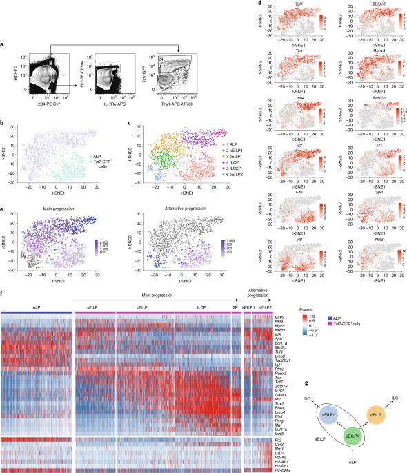
examined the transcriptional and functional heterogeneity of ILC progenitors, and studied the precursor–product relationships that link the subsets identified. This analysis identified two
successive stages of ILC development within T cell factor 1-positive (TCF-1+) early innate lymphoid progenitors (EILPs), which we named ‘specified EILPs’ and ‘committed EILPs’. Specified
EILPs generated dendritic cells, whereas this potential was greatly decreased in committed EILPs. TCF-1 was dispensable for the generation of specified EILPs, but required for the generation
of committed EILPs. TCF-1 used a pre-existing regulatory landscape established in upstream lymphoid precursors to bind chromatin in EILPs. Our results provide insight into the mechanisms by
which TCF-1 promotes developmental progression of ILC precursors, while constraining their dendritic cell lineage potential and enforcing commitment to ILC fate. Access through your
institution Buy or subscribe This is a preview of subscription content, access via your institution ACCESS OPTIONS Access through your institution Access Nature and 54 other Nature Portfolio
journals Get Nature+, our best-value online-access subscription $29.99 / 30 days cancel any time Learn more Subscribe to this journal Receive 12 print issues and online access $209.00 per
year only $17.42 per issue Learn more Buy this article * Purchase on SpringerLink * Instant access to full article PDF Buy now Prices may be subject to local taxes which are calculated
during checkout ADDITIONAL ACCESS OPTIONS: * Log in * Learn about institutional subscriptions * Read our FAQs * Contact customer support SIMILAR CONTENT BEING VIEWED BY OTHERS EFFECTOR
DIFFERENTIATION DOWNSTREAM OF LINEAGE COMMITMENT IN ILC1S IS DRIVEN BY HOBIT ACROSS TISSUES Article 30 August 2021 STAGE-SPECIFIC GATA3 INDUCTION PROMOTES ILC2 DEVELOPMENT AFTER LINEAGE
COMMITMENT Article Open access 05 July 2024 RECIPROCAL TRANSCRIPTION FACTOR NETWORKS GOVERN TISSUE-RESIDENT ILC3 SUBSET FUNCTION AND IDENTITY Article 23 September 2021 DATA AVAILABILITY The
accession number for the raw data of the RNA-Seq is GSE113767. The accession number for the raw data of the DNase-Seq and ChIC-Seq is GSE128483. All other relevant data are available from
the corresponding author on request. REFERENCES * Harly, C., Cam, M., Kaye, J. & Bhandoola, A. Development and differentiation of early innate lymphoid progenitors. _J. Exp. Med._ 215,
249–262 (2018). Article CAS Google Scholar * Jeevan-Raj, B. et al. The transcription factor Tcf1 contributes to normal NK cell development and function by limiting the expression of
granzymes. _Cell Rep._ 3, 613–626 (2017). Article Google Scholar * Weber, B. N. et al. A critical role for TCF-1 in T-lineage specification and differentiation. _Nature_ 476, 63–68 (2011).
Article CAS Google Scholar * Yang, Q. et al. TCF-1 upregulation identifies early innate lymphoid progenitors in the bone marrow. _Nat. Immunol._ 16, 1044–1050 (2015). Article CAS
Google Scholar * Hoyler, T. et al. The transcription factor GATA-3 controls cell fate and maintenance of type 2 innate lymphoid cells. _Immunity_ 37, 634–648 (2012). Article CAS Google
Scholar * Klose, C. S. et al. Differentiation of type 1 ILCs from a common progenitor to all helper-like innate lymphoid cell lineages. _Cell_ 157, 340–356 (2014). Article CAS Google
Scholar * Constantinides, M. G., McDonald, B. D., Verhoef, P. A. & Bendelac, A. A committed precursor to innate lymphoid cells. _Nature_ 508, 397–401 (2014). Article CAS Google
Scholar * Yu, Y. et al. Single-cell RNA-Seq identifies a PD-1hi ILC progenitor and defines its development pathway. _Nature_ 539, 102–106 (2016). Article CAS Google Scholar * Ji, Z.
& Ji, H. TSCAN: pseudo-time reconstruction and evaluation in single-cell RNA-Seq analysis. _Nucleic Acids Res._ 44, e117 (2016). Article Google Scholar * Murphy, T. L. et al.
Transcriptional control of dendritic cell development. _Annu. Rev. Immunol._ 34, 93–119 (2016). Article CAS Google Scholar * Grajales-Reyes, G. E. et al. Batf3 maintains autoactivation of
_Irf8_ for commitment of a CD8α+ conventional DC clonogenic progenitor. _Nat. Immunol._ 16, 708–717 (2015). Article CAS Google Scholar * Schlitzer, A. et al. Identification of cDC1- and
cDC2-committed DC progenitors reveals early lineage priming at the common DC progenitor stage in the bone marrow. _Nat. Immunol._ 16, 718–728 (2015). Article CAS Google Scholar * Heng, T.
S., Painter, M. W. and The Immunological Genome Project Consortium. The Immunological Genome Project: networks of gene expression in immune cells. _Nat. Immunol._ 9, 1091–1094 (2008). *
Schlenner, S. M. et al. Fate mapping reveals separate origins of T cells and myeloid lineages in the thymus. _Immunity_ 32, 426–436 (2010). Article CAS Google Scholar * Bell, J. J. &
Bhandoola, A. The earliest thymic progenitors for T cells possess myeloid lineage potential. _Nature_ 452, 764–767 (2008). Article CAS Google Scholar * Morrison, S. J., Wandycz, A. M.,
Hemmati, H. D., Wright, D. E. & Weissman, I. L. Identification of a lineage of multipotent hematopoietic progenitors. _Development_ 124, 1929–1939 (1997). CAS PubMed Google Scholar *
Specht, A. T. & Li, J. LEAP: constructing gene co-expression networks for single-cell RNA-sequencing data using pseudotime ordering. _Bioinformatics_ 33, 764–766 (2017). CAS PubMed
Google Scholar * Ku, W. L. et al. Single-cell chromatin immunocleavage sequencing (scChIC-seq) to profile histone modification. _Nat. Methods_ 16, 323–325 (2019). Article CAS Google
Scholar * Li, L. et al. A far downstream enhancer for murine Bcl11b controls its T-cell specific expression. _Blood_ 122, 902–911 (2013). Article CAS Google Scholar * Ohmura, S. et al.
Lineage-affiliated transcription factors bind the Gata3 Tce1 enhancer to mediate lineage-specific programs. _J. Clin. Invest._ 126, 865–878 (2016). Article Google Scholar * Sen, D. R. et
al. The epigenetic landscape of T cell exhaustion. _Science_ 354, 1165–1169 (2016). Article CAS Google Scholar * Heinz, S. et al. Simple combinations of lineage-determining transcription
factors prime cis-regulatory elements required for macrophage and B cell identities. _Mol. Cell_ 38, 576–589 (2010). Article CAS Google Scholar * Wang, D. et al. The transcription factor
Runx3 establishes chromatin accessibility of cis-regulatory landscapes that drive memory cytotoxic T lymphocyte formation. _Immunity_ 48, 659–674.e6 (2018). Article CAS Google Scholar *
Steinke, F. C. et al. TCF-1 and LEF-1 act upstream of Th-POK to promote the CD4+ T cell fate and interact with Runx3 to silence _Cd4_ in CD8+ T cells. _Nat. Immunol._ 15, 646–656 (2014).
Article CAS Google Scholar * Feyerabend, T. B. et al. Deletion of Notch1 converts pro-T cells to dendritic cells and promotes thymic B cells by cell-extrinsic and cell-intrinsic
mechanisms. _Immunity_ 30, 67–79 (2009). Article CAS Google Scholar * Wu, L. et al. Development of thymic and splenic dendritic cell populations from different hemopoietic precursors.
_Blood_ 98, 3376–3382 (2001). Article CAS Google Scholar * Shigematsu, H. et al. Plasmacytoid dendritic cells activate lymphoid-specific genetic programs irrespective of their cellular
origin. _Immunity_ 21, 43–53 (2004). Article CAS Google Scholar * Ishikawa, F. et al. The developmental program of human dendritic cells is operated independently of conventional myeloid
and lymphoid pathways. _Blood_ 110, 3591–3660 (2007). Article CAS Google Scholar * Rothenberg, E. V., Kueh, H. Y., Yui, M. A. & Zhang, J. A. Hematopoiesis and T-cell specification as
a model developmental system. _Immunol. Rev._ 271, 72–97 (2016). Article CAS Google Scholar * Farrell, J. A. et al. Single-cell reconstruction of developmental trajectories during
zebrafish embryogenesis. _Science_ 360, eaar3131 (2018). Article Google Scholar * Verbeek, S. et al. An HMG-box-containing T-cell factor required for thymocyte differentiation. _Nature_
374, 70–74 (1995). Article CAS Google Scholar * Aliahmad, P. & Kaye, J. Development of all CD4 T lineages requires nuclear factor TOX. _J. Exp. Med._ 205, 245–256 (2008). Article CAS
Google Scholar * Naik, S. H. et al. Development of plasmacytoid and conventional dendritic cell subtypes from single precursor cells derived in vitro and in vivo. _Nat. Immunol._ 8,
1217–1226 (2007). Article CAS Google Scholar * Onai, N. et al. Identification of clonogenic common Flt3+M-CSFR+ plasmacytoid and conventional dendritic cell progenitors in mouse bone
marrow. _Nat. Immunol._ 8, 1207–1216 (2007). Article CAS Google Scholar * Satija R., Butler A. & Hoffman P. Seurat: Tools for single cell fenomics. R package version 2.3.0
https://cran.r-project.org/web/packages/Seurat/index.html (2018). * Shannon, P. et al. Cytoscape: a software environment for integrated models of biomolecular interaction networks. _Genome
Res._ 13, 2498–2504 (2003). Article CAS Google Scholar * R Development Core Team _R: A Language and Environment for Statistical Computing_ (R Foundation for Statistical Computing, 2014).
* Dobin, A. et al. STAR: ultrafast universal RNA-Seq aligner. _Bioinformatics_ 29, 15–21 (2013). Article CAS Google Scholar * Liao, Y., Smyth, G. K. & Shi, W. The Subread aligner:
fast, accurate and scalable read mapping by seed-and-vote. _Nucleic Acids Res._ 41, e108 (2013). Article Google Scholar * Ritchie, M. E. et al. limma powers differential expression
analyses for RNA-sequencing and microarray studies. _Nucleic Acids Res._ 43, e47 (2015). Article Google Scholar * Leek, J. T. et al. sva: Surrogate variable analysis. R package version
3.26.0 (2017). * Cooper, J., Ding, Y., Song, J. & Zhao, K. Genome-wide mapping of DNase I hypersensitive sites in rare cell populations using single-cell DNase sequencing. _Nat. Protoc._
12, 2342–2354 (2017). Article CAS Google Scholar * Langmead, B. & Salzberg, S. L. Fast gapped-read alignment with Bowtie 2. _Nat. Methods_ 9, 357–359 (2012). Article CAS Google
Scholar * Zhang, Y. et al. Model-based analysis of ChIP-Seq (MACS). _Genome Biol._ 9, R137 (2008). Article Google Scholar * Tripathi, S. et al. Meta- and orthogonal integration of
influenza “OMICs” data defines a role for UBR4 in virus budding. _Cell Host Microbe_ 18, 723–735 (2015). Article CAS Google Scholar Download references ACKNOWLEDGEMENTS We thank H.-R.
Rodewald for sharing _Il7r-iCre_ mice and J. Richa from the Transgenic and Chimeric Mouse Facility, University of Pennsylvania, for injection of _Tcf7_YFP embryonic stem cells into mouse
blastocysts. We thank T. Ciucci, R. Bosselut, J. Chen, the CCR Sequencing Facility, the CCR Flow Cytometry Core Facility, and the DNA Sequencing Facility of the University of Pennsylvania
for technical support. This work utilized the computational resources of the NIH high-performance computing Biowulf cluster (http://hpc.nih.gov). This research was supported by the
Intramural Research Program of the NIH, National Cancer Institute and Center for Cancer Research, and by grants from the NIH (AI121080 and AI139874 to H.-H.X.), Veteran Affairs BLR&D
Merit Review Program (BX002903A to H.-H.X.), and Foundation pour la Recherche Médicale (DEQ20170839118 to C.H.) and National Research Agency Investissements d’Avenir via the program LabEX
IGO (ANR-11-LABX-0016-01 to C.H.). AUTHOR INFORMATION AUTHORS AND AFFILIATIONS * Laboratory of Genome Integrity, Center for Cancer Research, National Cancer Institute, National Institutes of
Health, Bethesda, MD, USA Christelle Harly, Devin Kenney & Avinash Bhandoola * CRCINA, INSERM, CNRS, Université d’Angers, Université de Nantes, Nantes, France Christelle Harly * LabEx
IGO ‘Immunotherapy, Graft, Oncology’, Nantes, France Christelle Harly * Systems Biology Center, National Heart, Lung, and Blood Institute, National Iinstitutes of Health, Bethesda, MD, USA
Gang Ren, Binbin Lai & Keji Zhao * Department of Medicine, Division of Translational Medicine and Human Genetics, Perelman School of Medicine, University of Pennsylvania, Philadelphia,
PL, USA Tobias Raabe * Department of Immunology and Microbial Disease, Albany Medical College, Albany, NY, USA Qi Yang * Office of Science and Technology Resources, Office of the Director,
Center for Cancer Research, National Cancer Institute, National Institutes of Health, Bethesda, MD, USA Margaret C. Cam * Department of Microbiology, Interdisciplinary Immunology Graduate
Program, Carver College of Medicine, University of Iowa, Iowa City, IA, USA Hai-Hui Xue * Iowa City Veterans Affairs Health Care System, Iowa City, IA, USA Hai-Hui Xue Authors * Christelle
Harly View author publications You can also search for this author inPubMed Google Scholar * Devin Kenney View author publications You can also search for this author inPubMed Google Scholar
* Gang Ren View author publications You can also search for this author inPubMed Google Scholar * Binbin Lai View author publications You can also search for this author inPubMed Google
Scholar * Tobias Raabe View author publications You can also search for this author inPubMed Google Scholar * Qi Yang View author publications You can also search for this author inPubMed
Google Scholar * Margaret C. Cam View author publications You can also search for this author inPubMed Google Scholar * Hai-Hui Xue View author publications You can also search for this
author inPubMed Google Scholar * Keji Zhao View author publications You can also search for this author inPubMed Google Scholar * Avinash Bhandoola View author publications You can also
search for this author inPubMed Google Scholar CONTRIBUTIONS C.H. designed the research and performed most of the experiments, alongside D.K. and G.R. C.H., B.L., M.C.C. and A.B. analyzed
the data. C.H., M.C.C., T.R. and A.B. produced the figures. C.H., T.R., Q.Y. and H.-H.X. designed and generated the new mouse models. C.H., K.Z. and A.B. directed and oversaw the
experiments. C.H. and A.B. wrote the paper. All authors helped to design the research, and read and commented on the manuscript. CORRESPONDING AUTHOR Correspondence to Avinash Bhandoola.
ETHICS DECLARATIONS COMPETING INTERESTS The authors declare no competing interests. ADDITIONAL INFORMATION PEER REVIEW INFORMATION: Ioana Visan was the primary editor on this article and
managed its editorial process and peer review in collaboration with the rest of the editorial team. PUBLISHER’S NOTE: Springer Nature remains neutral with regard to jurisdictional claims in
published maps and institutional affiliations. INTEGRATED SUPPLEMENTARY INFORMATION SUPPLEMENTARY FIGURE 1 REPRESENTATIVE GATING STRATEGY USED FOR FLOW CYTOMETRIC ANALYSIS. (A) Single cell
suspensions made from bone marrow are depleted of RBCs using osmotic lysis, then depleted for LinILC+ cells (see Methods), stained using DAPI, and analyzed by flow cytometry. (B) For
visualization of ILC precursors, an additional LinILC- Kit+ gate is applied before gating shown in Fig. 1a. SUPPLEMENTARY FIGURE 2 DIFFERENTIATION POTENTIAL OF EILPS _IN VITRO_. Flow
cytometric analysis of cultures from single EILPs sorted into 96 well plates and cultured for 10 days in either SF7-GM3 or SF7-GM3-MG6 conditions. (A-B) Examples of single EILP-derived
colonies showing the lineages identified. Arrows show successive gating. Numbers indicate the percentage of cells in each gate. (C) Composition of the ILC+ wells. Each column represents one
well, positive wells are indicated in black for individual lineage. (D) Quantification of absolute numbers of Mac-1+ cells per Mac-1+ colony in SF7-GM3 condition (n=50 colonies) and
SF7-GM3-MG6 condition (n=191 colonies). Data are presented as mean + SD. A two-tailed unpaired Student’s t-test was performed to determine significance, and showed that difference is not
significant. (E) Representative profile of Mac-1+ colonies gated on Mac-1+ cells. (F) Percentage of Mac-1+ wells containing cDC1, cDC2, or both lineages as shown in e. All data are
representative of three independent experiments. SUPPLEMENTARY FIGURE 3 GENERATION OF A _TCF7__YFP_ REPORTER MOUSE. (A) Strategy for the generation of a _Tcf7__YFP_ allele by insertion of a
_P2A-YFP_ sequence downstream of the _Tcf7_ C-terminus, followed by deletion of the floxed neo cassette using a _CMV-Cre_ mouse strain. The P2A ribosomal skipping peptide allows bicistronic
generation of separate _Tcf7_ and YFP molecules, but both driven by the _Tcf7_ promoter at equimolar amounts. _Tcf7_ exons are numbered (E3-E9), and the scale and the location of the MfeI
restriction sites and Southern blot probe used to screen the targeted ES clones are indicated. (B) Identification by Southern blot of an ES clone with the expected homologous recombination
(+/-) compared to a wild-type clone (-/-). Genomic DNA was extracted, digested with MfeI, and blotted with the probe shown in a. (C) Flow cytometric analysis of LinILC- Kit+ CD122low α4β7+
BM cells of the indicated mouse strains. Data are representative of three independent experiments. SUPPLEMENTARY FIGURE 4 CHARACTERIZATION OF EILP SUBSETS. (A) RNA-seq analysis of ALPs,
sEILPs, cEILPs and ILCPs. Hierarchical clustering using complete linkage calculated from Euclidian distances. (B-C) Flow cytometric analysis of cultures from single sEILPs and cEILPs sorted
into 96 well plates and cultured for 10 days in either SF7-GM3 or SF7-GM3-MG6 conditions. Data are pooled from 2 out of 3 representative experiments. (B) Frequency of wells containing ILC
progenitors as shown in Supplementary Fig. 2a, in SF7-GM3 condition. Data are presented as % of ILC positive wells ± SEP for n=78 sEILP wells and n=143 cEILP wells. (C) Frequency of ILC
positive colonies containing the indicated combination of mature ILCs, identified as shown in Supplementary Fig. 2a. (D) Flow cytometric analysis of sEILPs and cEILPs. (E) Flow cytometric
analysis of intracellular DAPI staining on sEILPs and cEILPs. (F) Flow cytometric analysis of cultures from sEILPs after two days in SF7 condition. Based on Mac-1, TCF-1, Thy1, and PLZF
expression (left), the derived populations were separated into DC (Mac-1+, grey), sEILP (TCF-1+ PLZF- Thy1-, black), cEILP (TCF-1+ PLZF+ Thy1-, orange), and ILCP (TCF-1+ PLZF+ Thy1+,
purple), and analyzed for expression of transcription factors (right). Arrows show successive gating. (G-H) Flow cytometric analysis of sEILP1s (green), sEILP2s (blue), cEILPs (orange) and
pre-DCs (grey) in _Tcf7__YFP_ mice (G) or wild-type mice (H). (I) Flow cytometric analysis of cultures from _Tcf7-_YFP+ sEILP1s and sEILP2s after two days in SF7 condition, gated on
_Tcf7-_YFP- cells. (J-K) Flow cytometric analysis of cultures from single sEILP1 and sEILP2 cells sorted into 96 well plates after 10 days in SF7-GM3 or SF7-GM3-MG6 conditions. (J)
Quantification of wells containing cDC1s, cDC2s, or both lineages as defined in Supplementary Fig. 2e. (K) Quantification of absolute numbers of DCs per DC positive colony in n=159 colonies
derived from sEILP1s and n=6 colonies derived from sEILP2s. Data are presented as mean + SD. A two-tailed unpaired Student’s t-test with Welsh correction was performed to determine
significance. ***_p_<0.005. (E,F,I) Numbers indicate the percentage of cells in each gate. All data are representative of three independent experiments. SUPPLEMENTARY FIGURE 5 DC
POTENTIAL OF EILPS _IN VIVO_. (A-B) Flow cytometric analysis of BM cells from wild-type mouse defining the indicated populations. Arrows show successive gating. (C) Flow cytometric analysis
of pre-DCs defined in a (grey), pre-cDC1s defined in b (blue), sEILPs (black) and CD11c+ EILPs defined in Fig. 4d (red). (D) Flow cytometric analysis of the indicated BM precursors (black)
compared to Lin Kit BM cells (grey). (E) Flow cytometric analysis of _Il7r-Cre R26-stop-YFP Tcf7__EGFP/+_ DC subsets defined in b. (A-E) Data are representative of three independent
experiments. (F) Flow cytometric analysis of BM cells from CD45.1+ mice that were lethally irradiated (850 rads), injected with _Tox__-/-_ or wild-type littermate CD45.2+LinILC-KithighSca-1+
BM cells mixed with CD45.1+LinILC-KithighSca-1+ BM cells, and reconstituted for 10–12 weeks. Profiles of LinILC-KithighSca-1+ cells and granulocytes from BM, and cDC1s (CD8α+Mac-1lo) and
cDC2s (CD8α-Mac-1hi) from spleen are shown. Data are pooled from two independent experiments that gave similar results, and presented as mean ± SEM for n=7 mice per group. A two-tailed
unpaired Student’s t-test was performed to determine significance. ns, not significant. (A,B,E,F) Numbers indicate the percentage of cells in each gate. SUPPLEMENTARY FIGURE 6 GENERATION OF
A _TCF7__EGFPNULL_ MOUSE AND ANALYSIS OF EILPS IN _TCF7__NULL_ MICE. (A) Strategy of generation of a _Tcf7__EGFPnull_ allele by breeding the _Tcf7__EGFP_ mouse with the _CMV-Cre_ mouse
strain. (B) Flow cytometric analysis of CD3ε+ splenocytes from _Tcf7__-/-_ (grey shaded histogram), _Tcf7__EGFP/+_ (black histogram), and _Tcf7__EGFPnull/-_ (red histogram) mice for GFP
expression on unfixed samples, or TCF-1 expression detected by intracellular staining with antibodies targeting either the N-terminal (C63D9) or C-terminal (C46C7) domains of TCF-1. (C) Flow
cytometric analysis of thymus from mice of the indicated genotype. (D) Numbers of thymocytes for _Tcf7__-/-_ (n=4), _Tcf7__EGFP/+_ (n=6), and _Tcf7__EGFPnull/-_ (n=6) mice pooled from three
independent experiments. Data are presented as mean ± SEM. (E) Flow cytometric analysis of LinILC- Kit+ CD122low BM cells from _Tcf7__EGFPnull/+_ and _Tcf7__EGFPnull/-_ littermate mice. (F)
Flow cytometric analysis of EILPs from e. (G) Flow cytometric analysis of LinILC-depleted BM cells from mice of the indicated genotype. Arrows show successive gating. (H) Flow cytometric
analysis and quantification of LinILC- Kit+ BM cells of _Tcf7__EGFPnull/+_ and _Tcf7__EGFPnull/-_ littermate mouse. Data are presented as mean ± SEM for n=3 mice per group. (C,E,G) Numbers
indicate the percentage of cells in each gate. (D,H) A two-tailed unpaired Student’s t-test was performed to determine significance. ns, not significant; **_p_<0.01, ***_p_<0.005.
(B-C,E-H) Data are representative of three independent experiments. (i) RNA-seq analysis averaged from 3 ALP samples, 2 _Tcf7__EGFPnull/-_ EILP samples, and 2 _Tcf7__EGFPnull/+_ sEILP
samples. Significance was calculated using a linear model (anova), applying the empirical Bayes method for estimating variance. Volcano plots show significantly upregulated or downregulated
genes by more than two-fold from ALPs to wild-type sEILPs colored in red and green respectively. SUPPLEMENTARY FIGURE 7 TCF-1 MECHANISM OF ACTION. (A,B) Flow cytometric analysis of cultures
from single sEILP sorted from _Tcf7__EGFPnull/+_ and _Tcf7__EGFPnull/-_ littermate mice into 96 well plates, after 10 days in SF7-GM3 or SF7-GM3-MG6 conditions. (A) Quantification of wells
containing ILCs, DCs, or both lineages. (B) Quantification of absolute numbers of Mac-1+ DCs per DC positive colony. Data are pooled from both cytokine conditions and presented as mean + SD.
Statistics are calculated using n=12 DC positive colonies derived from _Tcf7__EGFPnull/+_ sEILPs and n=34 DC positive colonies derived from _Tcf7__EGFPnull/-_ sEILPs. A two-tailed unpaired
Student’s t-test with Welsh correction was performed to determine significance. **_p_<0.01. (C-E) scRNA-seq analysis of _Tcf7_-GFP_+_ BM progenitors isolated from _Tcf7__EGFPnull/-_ mice
and compared to wild-type ALPs and _Tcf7_-GFP_+_ BM progenitors. (C) _t_-SNE plots showing pseudo-time ordering scores of individual ALP (n=786 cells), _Tcf7_-GFP_+_ _Tcf7__EGFP/+_ cells
(n=1799) and _Tcf7_-GFP_+_ _Tcf7__EGFPnull/-_ cells (n=594) shown in Fig. 6b along the two progressions from Fig. 1e. The ordering score of individual cells is represented in colors going
from light grey to violet for a given progression. Cells that are not part of the progression are dark grey. (D) Quantification of TCF-1 deficient and sufficient sEILP1s in each progression
from b, calculated as percentage of sEILP1. The size of the circle is relative to the percentage of cells. (E) Expression of the indicated genes of individual TCF-1 deficient (black) and
sufficient (pink) sEILP1s in each progression (main or alt. for alternative) as shown in c and d. Statistics are calculated on n=224 main sEILP1s and n=75 alt. sEILP1s that are TCF-1
deficient, and n=585 main sEILP1s and n=84 alt. sEILP1s that are TCF-1 sufficient. (F) LEAP analysis modelling the transcriptional network underlying ILC development. Interactions between
transcription factors. Only interactions with the 10 most connected controllers are represented. Controllers downregulated during ILC development are shown in green, controllers upregulated
are in red. The size of each controller is proportional to its network connectivity. See Supplementary Table 6 for the whole network. (G) TCF-1 ChIC-seq analysis in EILPs and DNase-seq
analysis in ALPs, EILPs, ILCPs. Heat map centered on TCF-1 binding sites in EILPs (± 2kb) that are not located in region of DNase I hypersensitivity in ALPs (left), and quantification of
DNase I hypersensitivity enrichment (right). (H) scRNA-seq analysis as in c-e. Expression of the indicated genes of individual TCF-1 deficient (n=224, black) and sufficient (n=585, pink)
sEILP1s from the Main developmental progression as shown in c and d. (E,H) A two-sided Wilcoxon rank-sum test was used to determine the significance of gene expression differences between
TCF-1 deficient and sufficient cells for a given subset. **_p_<0.01, ***_p_<0.005. See also Supplementary Table 4. (I) Scheme of early ILC development showing the progenitor-successor
relationships between EILP subsets, and their developmental fate. The transcription factors required for the described developmental progression are indicated. SUPPLEMENTARY INFORMATION
SUPPLEMENTARY INFORMATION Supplementary Figs. 1–7 REPORTING SUMMARY SUPPLEMENTARY TABLE 1: GENES DYNAMICALLY EXPRESSED DURING WILD-TYPE ILC DEVELOPMENT IN SCRNA-SEQ Cell clusters separated
by pseudotime were compared with each other. Clusters as defined in Fig. 1c are compared along the main (ALP (_n_ = 752), sEILP1 (_n_ = 585), cEILP (_n_ = 409), ILCP (_n_ = 414), ILC2P (_n_
= 111)) or alternative pseudotimes (ALP (_n_ = 751), sEILP1 (_n_ = 84), sEILP2 (_n_ = 187)), as defined in Fig. 1e. Log-transformed average expressions and fold changes are shown. A
two-sided Wilcoxon rank-sum test was used to determine significance. SUPPLEMENTARY TABLE 2: GENES DIFFERENTIALLY EXPRESSED BETWEEN TCF7EGFPNULL/− EILPS AND WILD-TYPE SEILPS IN BULK RNA-SEQ
Gene expression is shown as the log-transformed average for each sample. Fold changes in expression between TCF-1-deficient EILPs and wild-type sEILPs are indicated. SUPPLEMENTARY TABLE 3:
GENES DIFFERENTIALLY EXPRESSED BETWEEN TCF7EGFPNULL/− AND WILD-TYPE SEILP1S AND SEILP2S IN SCRNA-SEQ The gene expression of TCF-1-deficient and sufficient cells separated by clusters, as
shown in Fig. 6c, was compared. Statistics were calculated on _n_ = 270 sEILP1s and _n_ = 276 sEILP2s that were TCF-1 deficient, and _n_ = 615 sEILP1s and _n_ = 187 sEILP2s that were TCF-1
sufficient. Log-transformed average expressions and fold changes are shown. A two-sided Wilcoxon rank-sum test was used to determine significance. SUPPLEMENTARY TABLE 4: GENES DIFFERENTIALLY
EXPRESSED BETWEEN TCF7EGFPNULL/− AND WILD-TYPE SEILP1S SEPARATED BY PSEUDOTIMES IN SCRNA-SEQ The gene expression of TCF-1-deficient and sufficient sEILP1s separated by pseudotimes, as shown
in Supplementary Fig. 7c,d, was compared. Statistics were calculated on _n_ = 224 main sEILP1s and _n_ = 75 alternative sEILP1s that were TCF-1 deficient, and _n_ = 585 main sEILP1s and _n_
= 84 alternative sEILP1s that were TCF-1 sufficient. Log-transformed average expressions and fold changes are shown. A two-sided Wilcoxon rank-sum test was used to determine significance.
SUPPLEMENTARY TABLE 5: CORRELATION NETWORK Putative regulatory relationships between controllers and targets are indicated, along with their corresponding maximum absolute correlation
coefficient. All cells in the main trajectory of differentiation (_n_ = 2,633) were used to calculate Pearson’s correlations. Only maximum absolute correlations of ≥0.2 were considered,
which corresponded to an FDR < 5 × 10⁻⁵. The pseudotime lag of expression between a controller and its targets is indicated. SUPPLEMENTARY TABLE 6: TCF-1 CHIC-SEQ PEAKS The coordinates
for individual TCF-1 binding peaks, as identified by ChIC-Seq, are indicated and mapped to the nearest gene. SUPPLEMENTARY TABLE 7: TCF-1 GENE TARGETS TCF-1 gene targets were identified
using the correlation network (Supplementary Table 5), TCF-1 binding (Supplementary Table 6) and _Tcf7_EGFPnull/− RNA-Seq data (Supplementary Tables 3 and 4). RIGHTS AND PERMISSIONS Reprints
and permissions ABOUT THIS ARTICLE CITE THIS ARTICLE Harly, C., Kenney, D., Ren, G. _et al._ The transcription factor TCF-1 enforces commitment to the innate lymphoid cell lineage. _Nat
Immunol_ 20, 1150–1160 (2019). https://doi.org/10.1038/s41590-019-0445-7 Download citation * Received: 08 August 2018 * Accepted: 12 June 2019 * Published: 29 July 2019 * Issue Date:
September 2019 * DOI: https://doi.org/10.1038/s41590-019-0445-7 SHARE THIS ARTICLE Anyone you share the following link with will be able to read this content: Get shareable link Sorry, a
shareable link is not currently available for this article. Copy to clipboard Provided by the Springer Nature SharedIt content-sharing initiative