Play all audios:
First envisioned for determining crystalline structures, ptychography has become a useful imaging tool for microscopists. However, ptychography remains underused by biomedical researchers
due to its limited resolution and throughput in the visible light regime. Recent developments of spatial- and Fourier-domain ptychography have successfully addressed these issues and now
offer the potential for high-resolution, high-throughput optical imaging with minimal hardware modifications to existing microscopy setups, often providing an excellent trade-off between
resolution and field of view inherent to conventional imaging systems, giving biomedical researchers the best of both worlds. Here, we provide extensive information to enable the
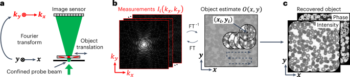
implementation of ptychography by biomedical researchers in the visible light regime. We first discuss the intrinsic connections between spatial-domain coded ptychography and Fourier
ptychography. A step-by-step guide then provides the user instructions for developing both systems with practical examples. In the spatial-domain implementation, we explain how a
large-scale, high-performance blood-cell lens can be made at negligible expense. In the Fourier-domain implementation, we explain how adding a low-cost light source to a regular microscope
can improve the resolution beyond the limit of the objective lens. The turnkey operation of these setups is suitable for use by professional research laboratories, as well as citizen
scientists. Users with basic experience in optics and programming can build the setups within a week. The do-it-yourself nature of the setups also allows these procedures to be implemented
in laboratory courses related to Fourier optics, biomedical instrumentation, digital image processing, robotics and capstone projects.
The main data supporting this study are available within the article, Supplementary Data and the primary supporting study10,11,14. Experimental datasets for both setups in this study are
available in Zenodo: https://doi.org/10.5281/zenodo.7492626.
All related MATLAB and Arduino code is provided in Supplementary Software. Additional code for testing experimental datasets is available in Zenodo: https://doi.org/10.5281/zenodo.7492626.
We thank Z. Bian and A. Pirhanov for their assistance in sample preparation. This work was partially supported by the UConn SPARK grant, UConn Research Excellence Program, National Science
Foundation award 2012140 and National Institute of Health award U01-NS113873. P.S. also acknowledges the support of the Thermo Fisher Scientific Fellowship.
These authors contributed equally: Shaowei Jiang, Pengming Song, Tianbo Wang.
Department of Biomedical Engineering, University of Connecticut, Storrs, USA
Shaowei Jiang, Pengming Song, Tianbo Wang, Liming Yang, Ruihai Wang, Chengfei Guo, Bin Feng & Guoan Zheng
Hangzhou Institute of Technology, Xidian University, Hangzhou, China
Department of Electronic and Electrical Engineering, University of Sheffield, Sheffield, UK
Diamond Light Source, Harwell Science and Innovation Campus, Chilton, UK
G.Z. conceived the project. S.J., P.S. and G.Z. designed the pipeline. S.J., P.S., T.W. and G.Z. developed the prototype systems and prepared the display items. S.J., P.S., T.W. and L.Y.
developed the data acquisition and processing pipelines for the protocol. T.W. and C.G. prepared all SolidWorks design files for the protocols. All authors contributed to the writing of the
manuscript.
G.Z. is a named inventor on the following patents related to Fourier ptychography (US Patent, nos. 9,817,224, 9,864,184, 9,497,379) and coded ptychography (US Patent, no. 11,487,099).
Nature Protocols thanks Zhengjun Liu, Fucai Zhang and the other, anonymous, reviewer(s) for their contribution to the peer review of this work.
Publisher’s note Springer Nature remains neutral with regard to jurisdictional claims in published maps and institutional affiliations.
Zheng, G. et al. Nat. Photonics 7, 739-745 (2013): https://doi.org/10.1038/nphoton.2013.187
Jiang, S. et al. ACS Photonics 8, 3261-3271 (2021): https://doi.org/10.1021/acsphotonics.1c01085
Jiang, S. et al. Biosens. Bioelectron. 196, 113699 (2022): https://doi.org/10.1016/j.bios.2021.113699
Springer Nature or its licensor (e.g. a society or other partner) holds exclusive rights to this article under a publishing agreement with the author(s) or other rightsholder(s); author
self-archiving of the accepted manuscript version of this article is solely governed by the terms of such publishing agreement and applicable law.
Anyone you share the following link with will be able to read this content: