Play all audios:
ABSTRACT Marek’s disease virus (MDV) is a preferred vector in the construction of recombinant vaccines. However, bivalent vaccine based on MDV that confers full protection against both very
virulent Marek’s and infectious bursal disease virus (IBDV) infections in chickens has not been produced. Here we developed a system utilizing overlapping fosmid DNAs transfection that
rescues an MDV type 1 (MDV1) vaccine strain. Using this system, we inserted the IBDV VP2 gene at MDV1 genome sites UL41, US10 and US2. The VP2 protein was stably expressed in the recombinant
MDV-infected cells and self-assembled into IBDV subviral particles. Insertion of the VP2 gene did not affect the replication phenotype of MDV in cell cultures, nor did it increase the
virulence of the MDV vaccine strain in chickens. After challenge with very virulent IBDV, r814US2VP2 conferred full protection, whereas r814UL41VP2 and r814US10VP2 provided partial or no
protection. All the three recombinant vaccines provided full protection against very virulent MDV challenge in chickens. These results demonstrated that r814US2VP2 could be used as a
promising bivalent vaccine against both Marek’s and infectious bursal diseases in chickens. SIMILAR CONTENT BEING VIEWED BY OTHERS EX VIVO RESCUE OF RECOMBINANT VERY VIRULENT IBDV USING A
RNA POLYMERASE II DRIVEN SYSTEM AND PRIMARY CHICKEN BURSAL CELLS Article Open access 06 August 2020 MASS VACCINATION WITH REASSORTMENT-IMPAIRED LIVE H9N2 AVIAN INFLUENZA VACCINE Article Open
access 03 August 2024 A CHIMERIC VACCINE DERIVED FROM AUSTRALIAN GENOTYPE IV JAPANESE ENCEPHALITIS VIRUS PROTECTS MICE FROM LETHAL CHALLENGE Article Open access 31 July 2024 INTRODUCTION
Marek’s disease (MD) is a neoplastic and neuropathic disease in chickens that was first reported by Joszef Marek over a century ago1. Marek’s disease virus (MDV) strains have three
serotypes: serotype 1 (MDV1) includes all the pathogenic strains and the attenuated strains of these viruses; serotype 2 (MDV2) includes naturally non-pathogenic strains; and serotype 3
(MDV3) is represented by turkey herpes virus (HVT)2. MDV1 remains the only neoplastic disease for which an effective vaccine has been used successfully and widely3. MDV has a large genome
which consists of a unique long (UL) region and a unique short (US) region, both flanked with repeat sequences. The MDV genome has several regions that are nonessential for viral replication
and therefore, suitable for the insertion of foreign genes, rendering the MDV1 vaccine strains a desirable live virus vector for expressing foreign genes4,5,6,7. Infectious bursal disease
(IBD) is an acute contagious immunosuppressive disease of young chickens caused by infectious bursal disease virus (IBDV)8. Since the discovery of the classic strains during the first
outbreak of IBD in 1957, antigenic variants and very virulent IBDV (vvIBDV) strains have emerged9,10, which represented new challenges for effective prevention of IBD. Since IBDV causes
disease in young chickens, early immunization is important for the prevention of the viral infection. However, with the high levels of circulating maternal antibodies, the immunity of
attenuated live vaccines of IBDV can be easily inhibited11. To overcome the maternal antibodies, medium virulent vaccines of IBDV were used, which induced better protection than the
attenuated vaccines, however, at a cost of inducing bursal damage and the failure of immunity of other poultry vaccines12. In addition, the virulence of live vaccines could be increased
after passages in chickens13. Therefore, it is necessary to develop safer and more efficacious vaccines to prevent vvIBDV infection. The strictly ordered part of the IBDV capsid is made
exclusively by VP214, which is the major protective antigen of IBDV and contains the epitopes that are responsible for eliciting neutralizing antibodies15. Considering its lower
susceptibility to maternal antibodies and good safety, recombinant MDV1 vaccines expressing VP2 would be more desirable than the conventional IBDV live vaccines. In the early 1970s, HVT
vaccine was firstly introduced in the field for the control of MD3. After a decade of use, very virulent MDV (vvMDV) strains emerged and began to break through the HVT protection, prompting
the introduction of a more effective bivalent vaccine that consisted of the MDV2 SB-1 strain plus HVT16. However, by the early 1990s, very virulent plus MDV (vv+MDV) strains began to emerge
and overcome protection provided by bivalent vaccines16. Currently, MD control was achieved by using the attenuated MDV1 vaccines such as CVI988 which are proving to be probably the only
effective vaccine against some of currently prevalent vv and vv+MDV strains. The attenuated MDV1 strain, 814, was introduced in China since 1980s; this strain has been widely used in China
as an important live vaccine for the prevention of high virulent MDV infection with a proven record of efficacy and safety17,18,19. Here, we demonstrate a system for generating the 814
strain by transfecting overlapping fosmid DNAs. Using this system, the IBDV VP2 gene was inserted at different sites in MDV1 genome. The recombinant virus r814US2VP2 containing VP2 gene at
US2 site confers full protection against vvMDV and vvIBDV infection in chickens. These results advance the development of efficient recombinant MDV vaccines and rapid manipulation of the
viral genome for basic research. RESULTS CONSTRUCTION OF AN OVERLAPPING FOSMID SYSTEM FOR MDV RECONSTITUTION The intact MDV genomic DNA (~170 kb) was purified from cells infected with strain
814 (Fig. S1A). After the genomic DNA was sheared and end-repaired, 36~48 kb fragments were purified from the agarose gels. These fragments were inserted into fosmid vector pCC1FOS,
generating an MDV fosmid library. The size of each DNA fragment was approximately 40 kb, based on results of a NotI digestion of the recombinant fosmids (Fig. S1B). After end sequencing, 24
fosmids with MDV genomic DNA insertions were selected for virus rescue; the size and location of these DNA fragments in the 814 genome are shown in Table S1. For virus rescue, 10 sets of
fosmid clones, each of which consisted of five- or six-fosmid combinations covering the entire MDV genome, were transfected into primary CEFs (Table S1). After five days of transfection, all
of the 10 sets of fosmid combination-transfected cells exhibited MDV1 typical cytopathic effects (CPE). The transfection of set 2 resulted in the highest percent of plaques, therefore, this
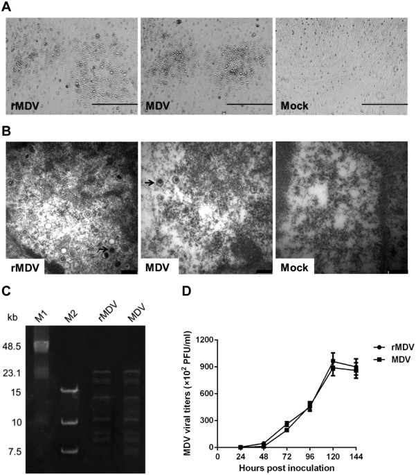
combination was used in further studies; virus rescued from this set was designated rMDV. CHARACTERIZATION OF THE RESCUED PARENTAL MDV VACCINE VIRUS The rescued parental vaccine virus rMDV
was characterized and compared with the wild-type virus. The rescued virus yielded plaques similar to those resulting from the wild-type virus (Fig. 1A), and generated in the nucleus
multiple naked virions indistinguishable in size and shape from those of wild-type virus (Fig. 1B). Pulsed field gel electrophoresis analysis of the rescued and wild-type vaccine virus DNA
digested with HindIII showed similar restriction patterns, indicating no rearrangement in the rescued viral DNA (Fig. 1C). _In vitro_ characterization of rMDV also showed that the growth
properties of this rescued virus were similar to those of the wild-type vaccine virus (Fig. 1D). These results indicate that we successfully rescued the MDV1 vaccine strain 814 using
overlapping fosmid DNAs, and that the rescued viruses had a genotype and replication phenotype similar to those of wild-type virus. GENERATION OF RECOMBINANT MDV CONTAINING IBDV VP2 GENE We
next inserted the VP2 gene of vvIBDV strain HLJ0504 into the UL41, US10 and US2 sites in the MDV1 vaccine genome for the construction of recombinant vaccines against IBDV. We firstly
constructed a VP2-expressing cassette by cloning the VP2 gene under the control of the Pec promoter, and then subcloned the cassette into an entry vector with the attL1 and attL2 sequences.
Finally, the VP2 expressing cassette was inserted into the above sites in the modified fosmids by LR reaction. The resultant fosmids containing _VP2_, 14-UL41VP2, 103-US10VP2, and 103-US2VP2
(Fig. 2B–D), were co-transfected with the other four parental fosmids for virus rescue. The MDV1-typical plaques observed in the CEFs and the virions detected in the electron microscopy
analysis indicated that recombinant MDVs expressing _VP2_ were rescued successfully (Fig. S2). BIOLOGICAL CHARACTERIZATION OF THE RECOMBINANT MDV EXPRESSING VP2 _VP2_ expression by the
recombinant MDVs was confirmed by the presence of green fluorescent signal in the infected cells, as detected by immunofluorescence (Fig. 3A). Western blot analysis of rMDV-VP2-infected CEF
lysate with anti-VP2 MAbs indicated that the molecular mass of the expressed VP2 was about 50 kDa (Fig. 3B). In both the immunofluorescence assay (IFA) and western blotting, r814UL41VP2
exhibited the strongest signal, followed by r814US2VP2; r814US10VP2 showed the weakest signal. VP2 expression was further quantified by fluorescence-activated cell-sorting (FACS); the
results revealed that r814UL41VP2 produced the highest fluorescent value and r814US10VP2 showed the lowest value, while r814US2VP2 produced VP2 at a medium level (Fig. 3C). Negative-stain
electron microscopy detected approximately 23 nm subviral IBDV particles in cells infected with r814UL41VP2 or r814US2VP2, but not in cells infected with r814US10VP2 or parental MDV (Fig.
3D). PCR amplification and sequencing of _VP2_ inserted in the recombinant MDVs passaged 20 times in CEFs confirmed that the recombinant MDVs had good genetic stability (Fig. 3E). VP2
expression from the serially passaged viruses was also confirmed by IFA with anti-VP2 MAbs (Fig. 3F). Further analysis demonstrated that the growth kinetics and magnitude of the recombinant
MDVs expressing _VP2_ were very similar to those of the parental virus (Fig. 3G), indicating that _VP2_ insertion in the MDV genome did not affect the replication of the MDV vaccine strain.
ANTIBODY RESPONSES AGAINST IBDV INDUCED BY RECOMBINANT MDVS IN CHICKENS We next tested the antibody responses against IBDV in specific-pathogen-free (SPF) chickens immunized with the
recombinant vaccines. As shown in Fig. 4A, r814UL41VP2 and r814US2VP2 induced detectable enzyme-linked immunosorbent assay (ELISA) antibodies against IBDV from 3 weeks post vaccination
(w.p.v), and the antibody titres induced by these two viruses before challenge at 4 w.p.v were comparable (_P_ > 0.05). By comparison, r814US10VP2 induced detectable antibodies after 4
w.p.v, and the antibody levels were lower than those induced by r814UL41VP2 and r814US2VP2 (_P_ < 0.05). Neutralizing antibodies against IBDV induced by the recombinant vaccines were
detected at 4 w.p.v (Fig. 4B). The results showed that r814UL41VP2 and r814US2VP2 induced neutralising antibodies at comparable levels (336 and 378, respectively), which were significantly
higher than that induced by r814US10VP2 (_P_ < 0.05). As expected, chickens immunized with the parental virus rMDV did not induce any detectable antibodies against IBDV during the
experiment. PROTECTIVE EFFICACY AGAINST LETHAL VVIBDV CHALLENGE IN CHICKENS To evaluate the protective efficacy of the recombinant viruses expressing VP2 against IBDV, vaccinated chickens
were challenged with vvIBDV HLJ0504 at 4 w.p.v. All the chickens vaccinated with r814UL41VP2 and r814US2VP2 were free of clinical signs and mortality after the challenge (Fig. 5A and B). In
contrast, all the chickens vaccinated with r814US10VP2 showed clinical signs of IBD, and died after challenge. Chickens immunized with the parental virus were fully susceptible to the IBDV
challenge, showing either high mortality (87%) or severe clinical signs (100%), whereas the healthy control group did not exhibit these signs. The bursa samples were collected after the
observation period, and the bursa: body weight index (BBIX) and histopathological bursal lesion score (HBLS) were calculated in order to analyse the extent of bursal atrophy and
histopathological lesions. As shown in Fig. 5C and D, all the chickens vaccinated with r814US2VP2 showed a BBIX value higher than 0.7 and a HBLS value no more than one, indicating that no
bursa had atrophy or gross lesions in this group. Among the 15 surviving chickens in the r814UL41VP2 group, two chickens exhibited bursal atrophy with a BBIX value lower than 0.7, and these
two chickens also showed mild bursal lesions (HBLS 2 and 3). The two surviving chickens in the rMDV group had severe bursal atrophy (BBIX 0.21 and 0.16) and gross lesions (HBLS 5), while no
bursal atrophy and lesions were observed in the healthy controls (0/10). Together the results of mortality, clinical signs and bursal lesions, vaccination with r814UL41VP2, r814US10VP2, and
r814US2VP2 conferred 87% (13/15), 0% (0/15), and 100% (15/15) protection against the lethal vvIBDV challenge. PROTECTIVE EFFICACY AGAINST LETHAL VVMDV CHALLENGE IN CHICKENS To determine if
the insertion of _VP2_ influenced the immunogenicity of the parental vaccine virus, chickens vaccinated with the recombinant MDVs as well as the parental vaccine virus were challenged with
the vvMDV Md5 strain 7 days after inoculation and examined for another 12 weeks. After challenge, 85% (17/20) of the chickens in the unvaccinated challenge control group died of MD, and the
remaining 3 chickens had histopathological MD lesions at the necropsy. The chickens vaccinated with the recombinant MDVs and the parental vaccine virus did not show clinical signs and had no
gross or histopathological tumors, indicating that the recombinant MDVs conferred full protection to chickens against vvMDV infection. DISCUSSION The development of recombinant vaccines has
been one of the most well studied aspects of molecular virology. MDV vaccine strain is a highly desirable live virus vector for expressing foreign antigen genes from other infectious
viruses. In practice, only MDV vaccines can be inoculated into day-old field chicks that have high titres of maternal antibodies, because the virus is transmitted cell to cell and is less
susceptible to maternal antibodies than some other virus vaccines3. Moreover, the MDV vaccines can induce lifetime immunity in chickens with just one vaccination7. In the previous studies,
recombinant HVT and MDV1 vaccines expressing antigen genes from several poultry viruses such as avian influenza virus7,20, Newcastle disease virus4,21, infectious bronchitis virus6 and
IBDV5,22, have been developed. However, with the emergence of vv and vv+MDV strains, HVT could no longer provide efficient protection against high virulent MDV infection23,24,25. By
comparison, attenuated MDV1 vaccines could induce more effective protection against the prevalent vv and vv+MDV strains, probably due to closer antigenic similarity with the oncogenic
strains3,16. From this point of view, attenuated MDV1 strains are more desirable than HVT as a viral vector for the development of bivalent recombinant vaccines against vvMDV infection and
other poultry diseases. In this study, three sites in the MDV1 vaccine genome were compared for the expression of _VP2_. The results revealed that the VP2 expression level from US2 was
significantly higher than that from US10. These results are consistent with those of a previous study, which demonstrated that the _HA_ gene from avian influenza virus exhibited a higher
expression level from US2 than from US10 in recombinant MDV7. Although several nonessential genes for viral growth have been identified in MDV1 UL area, there are no reports of recombinant
MDV1 containing foreign antigen genes in this area. In the present study, the VP2 gene was inserted into the UL41 gene without influencing the viral growth. The VP2 expression level from
UL41 was shown to be significantly higher than those from the US2 and US10 sites. By electron microscopy analysis, the subviral IBDV particles (SVPs) formed by VP2 were detected in the cells
infected with r814UL41VP2 or r814US2VP2, but not in cells inoculated with r814US10VP2, indicating that large amount of VP2 is necessary for the SVPs formation in cells. Animal experiments
indicated that the protective efficacy of the recombinant vaccines is influenced by the insertion site notably. The antibody responses induced by r814UL41VP2 and r814US2VP2 were comparable,
and were significantly higher than that induced by r814US10VP2. After challenge, r814US10VP2 did not provide any protection against mortality. The r814UL41VP2 fully protected against
clinical symptoms and mortality, but conferred partial protection against bursal atrophy and lesions. In contrast, r814US2VP2 provided full protection, not only against the development of
clinical signs and mortality, but also against bursal damage. The ineffective protection induced by r814US10VP2 could be due to the low expression of VP2, which was not sufficient for
inducing a protective immune response against IBDV. However, why r814UL41VP2, which expressed VP2 at the highest level and replicated similarly _in vitro_ with the parental virus, induced a
lower level of protection than r814US2VP2, needs to be further studied. The results suggested that the antigen expression level was not the only factor that influences the protective
efficacy of recombinant MDV vaccines, and other parameters, mainly the replication process of the virus _in vivo_, should be taken into consideration. With regard to developing a recombinant
MDV1 vaccine against IBDV, the US2 site, which expressed foreign genes at a medium level, might be better than the high-expression UL41 site and the low-expression US10 site. Whether this
is the case for generating recombinant MDV1 expressing other antigens needs to be determined, since the antigen characteristics and protective immunity are different among viruses.
Considering the uniquely advantageous characteristics of the attenuated MDV1 vector in the construction of recombinant vaccines against IBDV and its efficacious protection against high
virulent MDV, many MDV1-vectored recombinant vaccines expressing the VP2 gene of IBDV have been constructed and evaluated during the last two decades5,26. However, attempts to elicit
complete protection against vvIBDV infection in chickens using recombinant MDV1 vaccines have not been successful. Previously reported recombinant viruses expressing VP2 could hardly protect
against bursal lesions when challenged with vvIBDV, although they protected chickens against mortality. Tsukamoto _et al_. produced a recombinant MDV1 virus expressing VP2 using the CVI988
vaccine strain as the vector; this recombinant CVI988 protected chickens against vvIBDV-induced clinical signs and mortality, but only 55% chickens were protected against bursal lesions5.
Zhou _et al_. developed a recombinant CVI988 vaccine expressing IBDV VP2, which conferred 73% protection against vvIBDV challenge in SPF chickens and 87% protection in commercial chickens,
even with a high vaccination dose of 104 PFU per chicken26. In our study, we constructed a recombinant MDV1 expressing IBDV VP2 using a Chinese MDV1 vaccine strain, 814, as the viral vector.
By cloning the VP2 gene under control of a strong promoter and inserting the VP2 cassette into the US2 gene, the r814US2VP2 vaccine fully protected chickens not only against developing
clinical signs and mortality but also against the formation of bursal lesions when challenged with vvIBDV. The virulence of the HLJ0504 strain we used in the challenge experiment was
previously shown to be notably higher than that of the European reference vvIBDV strains27. After challenged with HLJ0504, 87% chickens died in the unvaccinated challenge group; the
mortality was higher than that reported in the previous studies5,26, indicating a higher pathogenic vvIBDV strain might be used here. Tsukamoto _et al_. constructed a recombinant HVT vaccine
expressing VP2, which was shown to be capable of inducing complete protection against lethal IBDV challenge22. However, HVT vector is inferior to the attenuated MDV1 vaccine strains
considering its limited protection against the prevalent high virulent MDV strains23,24,25. Since the attenuated MDV1 vaccine strain 814 was used here as the vector expressing VP2, the
r814US2VP2 also exhibits full protection against vvMDV lethal challenge. The results indicate that a more efficacious MDV1-vectored bivalent vaccine against both vvMDV and vvIBDV infection
in chickens was constructed in this study. Over the last two decades, much attention has been paid to MDV-vectored vaccines4,5,6,7. However, recombinant MDVs have been constructed
exclusively using traditional homologous recombination24 or the bacterial artificial chromosome (BAC) system28,29. These methods require either several rounds of plaque purification or the
time-consuming excision of the BAC sequence30,31. In a methodological improvement, Reddy _et al_. developed a cosmid-based system for the rescue of vvMDV Md5 strain32. However, the cosmid
system has not been used to construct recombinant MDV vaccines; only the generation of Md5 has been reported. The unwieldiness of the cosmid system may be due to the instability of the
cosmid clones containing large DNA inserts during routine maintenance and propagation33. To overcome the above limitations, fosmid, which is derived from the bacterial F episome and is
present in single or low copy number per cell, was introduced as a substitute for the conventional cosmid in genome library construction for its better stability and inducible copy number33.
The fosmid technology was also used in the construction of recombinant duck enteritis virus expressing the HA gene of avian influenza virus with high efficiency34. Here, the complete genome
of MDV 814 strain was cloned as overlapping fosmid DNAs, and the rescued virus was proven infectious with good immunogenicity. We next modified the fosmids by inserting a dual selection
marker flanked by the attR1 and attR2 sequences into three different sites in MDV genome, and constructed a versatile entry vector with a multiple clone sequence flanked by the attL1 and
attL2 sequences. With this system, we could generate the recombinant MDVs within two weeks after transfection without the need for repeated rounds of plaque purification and introduction of
marker genes. Hence, this system is more efficient than the traditional methods. This technology is highly valuable for constructing recombinant MDVs against viruses that easily undergo
antigenic drift, as vaccine update would be easier and more feasible. With the effective protection, the r814US2VP2 vaccine represents a promising bivalent vaccine against vvIBDV and vvMDV,
two lethal and immunosuppressive pathogens. Further studies are worthwhile to be conducted before its ready to use in the field farms such as the required vaccination dose, the required
level of immune against IBDV and the protective efficacy in commercial chickens with maternal antibodies. Furthermore, the establishment of an efficient fosmid rescue system for generating
recombinant MDVs should facilitate the analyses of MDV functional genes and vaccine-induced protective immunity against the neoplastic disease. MATERIAL AND METHODS ETHICS STATEMENT All
animal experiments were approved by the ethical review board of Harbin Veterinary Research Institute (HVRI) of the Chinese Academy of Agricultural Sciences (CAAS) and performed in accordance
with approved animal care guidelines and protocols. The animal Ethics Committee approval number is SYXK (Heilongjiang) 2011022. VIRUSES AND CELLS MDV1 vaccine strain 81418 was used as the
parent virus for producing recombinant MDVs. The vvMDV Md535 and vvIBDV HLJ050427 strains were used as challenge viruses. MDVs were propagated in CEFs prepared from 10-day-old
specific-pathogen-free (SPF) chicken embryos. CONSTRUCTION OF THE MDV FOSMID LIBRARY MDV strain 814 was propagated in CEFs until 80–90% CPE was obtained. The viral DNA was purified from
infected cells using hypotonic lysis to release virus particles followed by micrococcal nuclease treatment to degrade cellular DNA36. An MDV fosmid library was constructed using the Copy
Control Fosmid Library Production Kit (Epicentre). The viral DNA was sheared into 25~50 kb fragments. After blunt treatment and phosphorylation of the ends, the viral DNA was selected for
36–48 kb fragments with pulsed field gel electrophoresis (PFGE, Bio-Rad). The collected fragments were subsequently inserted into the cloning-ready fosmid vector pCC1FOS. The recombinant
fosmids were packaged using MaxPlax Lambda Packaging Extracts and plated on EPI300-T1 plating cells (Epicentre). The presence of the DNA fragment inserts in these fosmids was confirmed by
end sequencing with a pair of specific primers (5′-GGA TGT GCT GCA AGG CGA TTA AGT TGG-3′ and 5′-CTC GTA TGT TGT GTG GAA TTG TGA GC-3′). CONSTRUCTION OF RECOMBINANT FOSMIDS CONTAINING VP2
GENE Five fosmids (195, 214, 14, 96, and 103), which contain sequences spanning the entire genome of MDV1 strain 814, were selected for rescuing the parental virus (Fig. 2A). To facilitate
the insertion of foreign genes into MDV genome, the fosmids were modified by inserting a dual selected maker open reading frame encoding the kanamycin resistance (_KanR_) gene and the toxin
_ccdB_ gene, flanked by the attR1 and attR2 sequences, into the MDV genome at the UL41, US10 and US2 sites. The _KanR_ and ccdB-attR2 sequences were amplified from pMOD6 and pDEST22 plasmid.
The aatR1-KanR-ccdB-aatR2 cassette was then combined by overlapping PCR, and 50 nucleotides matching the sequences of different areas of UL41, US10, and US2 of the 814 strain genome were
introduced into the two ends of the cassette. The resultant cassettes with homology arms were inserted into 14 and 103 with Counter-Selection BAC modification kit (Gene Bridges). To simplify
the insertion of foreign genes into MDV genome, a versatile entry vector pENTR-mcs was constructed by replacing the _gus_ gene flanked by attL1 and attL2 in the plasmid pENTR-gus
(Invitrogen) with a multiple cloning sequence. For _VP2_ expression, _VP2_ from the vvIBDV strain HLJ0504 was cloned into pCAGGS plasmid to form the VP2-expressing cassette. The VP2 cassette
was then inserted into pENTR-mcs. To insert the VP2 cassette into the desired sites in MDV1 genome, pENTR-VP2 was mixed with the modified fosmids and treated with LR Clonase II enzyme
(Invitrogen). The mixtures were transformed into _Escherichia coli_ EPI300-T1 cells. RESCUE AND CHARACTERIZATION OF THE RECOMBINANT MDVS The five fosmids with or without VP2 insertions that
covered the entire MDV genome were used for virus rescue. Viral DNA inserts were released from purified fosmids by digestion with NotI and purified before transfection. Two micrograms of
each fosmid DNA were used to transfect primary CEFs in 60-mm dishes by using the calcium phosphate procedure37. The cytopathic effects (CPE)-positive samples were harvested and characterized
by electron microscopy. To verify that the VP2 gene was inserted at the desired sites, viral genomic DNAs were isolated and analysed by PCR and sequencing. CHARACTERIZATION OF RESCUED MDVS
CEF cells infected with parental or recombinant MDVs were first characterized with electron microscopy after CPE appeared. Genomic DNA was extracted from the infected cells and digested with
HindIII for pulsed-field gel electrophoresis analyses. To verify that the VP2 gene was inserted into MDV genome at the desired site, genomic DNA was isolated from the rescued viruses and
analyzed by PCR with the forward primer specific to VP2 and the reverse primer matching US2. The PCR products were purified and sequenced. CONFIRMATION OF VP2 GENE EXPRESSION The expression
of VP2 from the recombinant MDVs was confirmed by IFA and western blotting with anti-VP2 MAb as the primary antibody and goat anti-mouse IgG conjugated with fluorescein isothiocyanate (FITC)
or DyLight 800 as the secondary antibody. To compare the VP2 expression levels, CEF cells were infected with 1,000 PFU of the recombinant MDVs and maintained for 120 h before harvesting.
After fixed and perforated, the cells were incubated first with mouse anti-VP2 MAb and then with FITC-conjugated goat anti-mouse IgG antibody. The mean fluorescence intensity (MFI) of
VP2-positive cells (10,000 events were captured per sample) was assessed by FACS analysis (FACSAria; Becton Dickinson). To determine whether VP2 expressed by the recombinant MDV
self-assembled into SVPs, negative-stain electron microscopy was performed as previously described38. Infected cell lysates were negatively stained with a 2% uranyl acetate aqueous solution,
and observed using a HITACHI H-7650 electron microscope. Digital images were processed using the iTEM software (Munster, Germany). STABILITY AND GROWTH PROPERTIES OF THE RESCUED VIRUSES To
evaluate the genetic stability of the recombinant viruses, viruses were passaged in CEFs 20 times. The inserted VP2 gene was detected by using PCR and sequencing. VP2 expression was
confirmed with fluorescence assays as described above. To investigate the growth properties of the recombinant MDVs, cells cultured in 6-well plates were inoculated with 100 PFU of different
viruses and harvested at different time points. The serial dilutions were inoculated onto fresh CEFs seeded on 6-well plates; plaques of the different dilutions were counted five days
later. PROTECTION AGAINST VVIBDV CHALLENGE To evaluate the protective efficacy of the recombinant viruses against vvIBDV, each of 15 1-day-old SPF chickens were vaccinated with 5,000 PFU of
the recombinant MDVs or the parental virus, and challenged with 105 EID50 of vvIBDV HLJ0504 at 28 days post vaccination. The clinical signs were quantified using a previously described
symptomatic index27. The dead and surviving chickens were evaluated for bursal atrophy with the bursa: body-weight index (BBIX)38. Bursae with a BBIX lower than 0.70 were considered
atrophied39. The bursae were further examined histopathologically and recorded using the histopathological bursal lesion score (HBLS)40. A HBLS value of no more than 1 (no or slight lesion)
was defined as protected against IBDV challenge. SEROLOGICAL TESTS The serum samples collected weekly were tested by ELISA (IDEXX, Westbrook, Maine). For virus neutralization (VN) assay,
triplicates of serum samples were diluted serially by two-fold and mixed with equal volume of 100 TCID50 of cell culture-adapted HLJ0504 virus. After 60 min of incubation, the mixtures were
added to CEFs followed by further incubation for 3 days. The VN titre was determined as the highest dilution at which there were no visible cytopathic effects. PROTECTION AGAINST VVMDV
CHALLENGE Each of 20 1-day-old SPF chickens was vaccinated subcutaneously with 2,000 PFU of the recombinant MDVs, and challenged intraperitoneally 7 days later with 1,000 PFU of the vvMDV
strain Md5. The chickens were examined for clinical signs and mortality for 12 weeks after the challenge. Both dead and surviving chickens were subjected to gross and histopathological
observations for MD lesions in the liver, kidneys, spleen, nerves, and skin. The protection index (PI) was calculated as described previously25. STATISTICAL ANALYSIS All data were presented
as the mean ± standard deviation (S.D.). One-way ANOVA was employed to evaluate the statistical differences among groups using SPSS 17.0 (SPSS Inc., Chicago, IL). Statistical significance
was set at _P_ < 0.05 for all tests. ADDITIONAL INFORMATION HOW TO CITE THIS ARTICLE: Li, K. _et al_. Recombinant Marek’s disease virus type 1 provides full protection against very
virulent Marek’s and infectious bursal disease viruses in chickens. _Sci. Rep._ 6, 39263; doi: 10.1038/srep39263 (2016). PUBLISHER'S NOTE: Springer Nature remains neutral with regard to
jurisdictional claims in published maps and institutional affiliations. REFERENCES * Marek, J. Multiple Nervenentzündung (Polyneuritis) bei Hühnern. Dtsch. Tierärztl. Wochenschr. 15,
417–421 (1907). Google Scholar * Biggs, P. M. & Nair, V. The long view: 40 years of Marek’s disease research and avian pathology. Avian Pathol. 41, 3–9 (2012). Article Google Scholar
* Venugopal, K. Marek’s disease: an update on oncogenic mechanisms and control. Res. Vet. Sci. 69, 17–23 (2000). Article CAS Google Scholar * Sakaguchi, M. et al. Protection of chickens
with or without maternal antibodies against both Marek’s and Newcastle diseases by one-time vaccination with recombinant vaccine of Marek’s disease virus type 1. Vaccine 16, 472–479 (1998).
Article CAS Google Scholar * Tsukamoto, K. et al. Protection of chickens against very virulent infectious bursal disease virus (IBDV) and Marek’s disease virus (MDV) with a recombinant
MDV expressing IBDV VP2. Virology 257, 352–362 (1999). Article CAS Google Scholar * Zhang, X., Wu, Y., Huang, Y. & Liu, X. Protection conferred by a recombinant Marek’s disease virus
that expresses the spike protein from infectious bronchitis virus in specific pathogen-free chicken. Virol. J. 9, 85 (2012). Article Google Scholar * Zhang, F. et al. Transcriptional
activity comparison of different sites in recombinant Marek’s disease virus for the expression of the H9N2 avian influenza virus hemagglutinin gene. J. Virol. Methods 207, 138–145 (2014).
Article CAS Google Scholar * van den Berg, T. P. Acute infectious bursal disease in poultry: a review. Avian Pathol. 29, 175–194 (2000). Article CAS Google Scholar * Jackwood, D. H.
& Saif, Y. M. Antigenic diversity of infectious bursal disease viruses. Avian Dis. 31, 766–770 (1987). Article CAS Google Scholar * Chettle, N., Stuart, J. C. & Wyeth, P. J.
Outbreak of virulent infectious bursal disease in East Anglia. Vet. Rec. 125, 271–272 (1989). Article CAS Google Scholar * Hsieh, M. K., Wu, C. C. & Lin, T. L. DNA-mediated
vaccination conferring protection against infectious bursal disease in broiler chickens in the presence of maternal antibody. Vaccine 28, 3936–3943 (2010). Article CAS Google Scholar *
Giambrone, J. J. et al. Effect of infectious bursal agent on the response of chickens to Newcastle disease and Marek’s disease vaccination. Avian Dis. 20, 534–544 (1976). Article CAS
Google Scholar * Muskett, J. C., Reed, N. E. & Thornton, D. H. Increased virulence of an infectious bursal disease live virus vaccine after passage in chicks. Vaccine 3, 309–312 (1985).
Article CAS Google Scholar * Coulibaly, F. et al. The birnavirus crystal structure reveals structural relationships among icosahedral viruses. Cell 120, 761–772 (2005). Article CAS
Google Scholar * Fahey, K. J., Erny, K. & Crooks, J. A conformational immunogen on VP2 of infectious bursal disease virus that induces virus-neutralizing antibodies that passively
protect chickens. J. Gen. Virol. 70, 1473–1481 (1989). Article CAS Google Scholar * Haq, K., Schat, K. A. & Sharif, S. Immunity to Marek’s disease: where are we now? Dev. Comp.
Immunol. 41, 439–446 (2013). Article CAS Google Scholar * Cui, H. Y. et al. Construction of an infectious Marek’s disease virus bacterial artificial chromosome and characterization of
protection induced in chickens. J. Virol. Methods 156, 66–72 (2009). Article CAS Google Scholar * Zhang, F. et al. Comparative full-length sequence analysis of Marek’s disease virus
vaccine strain 814. Arch. Virol. 157, 177–183 (2012). Article CAS Google Scholar * Cui, H. et al. Avirulent Marek’s disease virus type 1 strain 814 vectored vaccine expressing avian
influenza (AI) virus H5 haemagglutinin induced better protection than turkey herpesvirus vectored AI vaccine. PLoS One 8, e53340 (2013). Article ADS CAS Google Scholar * Li, Y. et al.
Recombinant herpesvirus of turkeys as a vector-based vaccine against highly pathogenic H7N1 avian influenza and Marek’s disease. Vaccine 29, 8257–8266 (2011). Article CAS Google Scholar *
Reddy, S. K. et al. Protective efficacy of a recombinant herpesvirus of turkeys as an in ovo vaccine against Newcastle and Marek’s diseases in specific-pathogen-free chickens. Vaccine 14,
469–477 (1996). Article CAS Google Scholar * Tsukamoto, K. et al. Complete, long-lasting protection against lethal infectious bursal disease virus challenge by a single vaccination with
an avian herpesvirus vector expressing VP2 antigens. J. Virol. 76, 5637–5645 (2002). Article CAS Google Scholar * Bülow, V. & Biggs, P. M. Differentiation between strains of Marek’s
disease virus and turkey herpesvirus by immunofluorescenece assays. Avian Pathol. 4, 133–146 (1975). Article Google Scholar * Darteil, R. et al. Herpesvirus of turkey recombinant viruses
expressing infectious bursal disease virus (IBDV) VP2 immunogen induces protection against an IBDV virulent challenge in chickens. Virology 211, 481–490 (1995). Article CAS Google Scholar
* Sonoda, K. et al. Development of an effective polyvalent vaccine against both Marek’s and Newcastle diseases based on recombinant Marek’s disease virus type 1 in commercial chickens with
maternal antibodies. J. Virol. 74, 3217–3226 (2000). Article CAS Google Scholar * Zhou, X. et al. Protection of chickens, with or without maternal antibodies, against IBDV infection by a
recombinant IBDV-VP2 protein. Vaccine 28, 3990–3996 (2010). Article CAS Google Scholar * Li, K. et al. Genetic, antigenic and pathogenic characterization of four infectious bursal
disease virus isolates from China suggests continued evolution of very virulent viruses. Infect. Genet. Evol. 30, 120–127 (2015). Article Google Scholar * Petherbridge, L. et al.
Replication-competent bacterial artificial chromosomes of Marek’s disease virus: novel tools for generation of molecularly defined herpesvirus vaccines. J. Virol. 77, 8712–8718 (2003).
Article CAS Google Scholar * Baigent, S. J. et al. Herpesvirus of turkey reconstituted from bacterial artificial chromosome clones induces protection against Marek’s disease. J. Gen.
Virol. 87, 769–776 (2006). Article CAS Google Scholar * Wagner, M., Jonjic, S., Koszinowski, U. H. & Messerle, M. Systematic excision of vector sequences from the BAC-cloned
herpesvirus genome during virus reconstitution. J. Virol. 73, 7056–7060 (1999). CAS PubMed PubMed Central Google Scholar * Zhao, Y., Petherbridge, L., Smith, L. P., Baigent, S. &
Nair, V. Self-excision of the BAC sequences from the recombinant Marek’s disease virus genome increases replication and pathogenicity. Virol. J. 5, 19 (2008). Article Google Scholar *
Reddy, S. M. et al. Rescue of a pathogenic Marek’s disease virus with overlapping cosmid DNAs: use of a pp38 mutant to validate the technology for the study of gene function. Proc. Natl.
Acad. Sci. USA 99, 7054–7059 (2002). Article ADS CAS Google Scholar * Kim, U. J., Shizuya, H., de Jong, P. J., Birren, B. & Simon, M. I. Stable propagation of cosmid sized human DNA
inserts in an F factor based vector. Nucleic Acids Res. 20, 1083–1085 (1992). Article CAS Google Scholar * Liu, J. et al. A duck enteritis virus-vectored bivalent live vaccine provides
fast and complete protection against H5N1 avian influenza virus infection in ducks. J. Virol. 85, 10989–10998 (2011). Article CAS Google Scholar * Tulman, E. R. et al. The genome of a
very virulent Marek’s disease virus. J. Virol. 74, 7980–7988 (2000). Article CAS Google Scholar * Volkening, J. D. & Spatz, S. J. Purification of DNA from the cell-associated
herpesvirus Marek’s disease virus for 454 pyrosequencing using micrococcal nuclease digestion and polyethylene glycol precipitation. J. Virol. Methods 157, 55–61 (2009). Article CAS Google
Scholar * Morgan, R. W., Cantello, J. L. & McDermott, C. H. Transfection of chicken embryo fibroblasts with Marek’s disease virus DNA. Avian Dis. 34, 345–351 (1990). Article CAS
Google Scholar * Dubochet, J. et al. Cryo-electron microscopy of vitrified specimens. Q. Rev. Biophys. 21, 129–228 (1988). Article CAS Google Scholar * Li, K. et al. Codon optimization
and woodchuck hepatitis virus posttranscriptional regulatory element enhance the immune responses of DNA vaccines against infectious bursal disease virus in chickens. Virus Res. 175, 120–127
(2013). Article CAS Google Scholar * Lucio, B. & Hitcher, B. Immunosuppression and active response induced by infectious bursal disease virus in chickens with passive antibodies.
Avian Dis. 24, 189–196 (1979). Article Google Scholar Download references ACKNOWLEDGEMENTS This research was supported by the grant from i) National “Twelfth Five-Year” Plan for Science
and Technology Support (2015BAD12B03); ii) the Major Project of National Natural Science Foundation of China (31430087); iii) Natural Science Foundation of Heilongjiang, China (QC2016042);
and iv) China Postdoctoral Science Foundation funded project (2015M580157). AUTHOR INFORMATION AUTHORS AND AFFILIATIONS * Avian Immunosuppressive Diseases Division, State Key Laboratory of
Veterinary Biotechnology, Harbin Veterinary Research Institute, Chinese Academy of Agricultural Sciences, Harbin, 150069, P.R. China Kai Li, Yongzhen Liu, Changjun Liu, Li Gao, Yanping
Zhang, Hongyu Cui, Yulong Gao, Xiaole Qi, Li Zhong & Xiaomei Wang Authors * Kai Li View author publications You can also search for this author inPubMed Google Scholar * Yongzhen Liu
View author publications You can also search for this author inPubMed Google Scholar * Changjun Liu View author publications You can also search for this author inPubMed Google Scholar * Li
Gao View author publications You can also search for this author inPubMed Google Scholar * Yanping Zhang View author publications You can also search for this author inPubMed Google Scholar
* Hongyu Cui View author publications You can also search for this author inPubMed Google Scholar * Yulong Gao View author publications You can also search for this author inPubMed Google
Scholar * Xiaole Qi View author publications You can also search for this author inPubMed Google Scholar * Li Zhong View author publications You can also search for this author inPubMed
Google Scholar * Xiaomei Wang View author publications You can also search for this author inPubMed Google Scholar CONTRIBUTIONS K.L. and X.W. conceived and designed the research; K.L. and
Y.L. performed the experiments; L.G. and L.Z. analyzed the data of experiments; C.L., Y.Z. and H.C. contributed with essential information of techniques; K.L. wrote the paper; Y.G. and X.Q.
were involved in the interpretation of the results and critically read the manuscript. ETHICS DECLARATIONS COMPETING INTERESTS The authors declare no competing financial interests.
ELECTRONIC SUPPLEMENTARY MATERIAL SUPPLEMENTARY INFORMATION RIGHTS AND PERMISSIONS This work is licensed under a Creative Commons Attribution 4.0 International License. The images or other
third party material in this article are included in the article’s Creative Commons license, unless indicated otherwise in the credit line; if the material is not included under the Creative
Commons license, users will need to obtain permission from the license holder to reproduce the material. To view a copy of this license, visit http://creativecommons.org/licenses/by/4.0/
Reprints and permissions ABOUT THIS ARTICLE CITE THIS ARTICLE Li, K., Liu, Y., Liu, C. _et al._ Recombinant Marek’s disease virus type 1 provides full protection against very virulent
Marek’s and infectious bursal disease viruses in chickens. _Sci Rep_ 6, 39263 (2016). https://doi.org/10.1038/srep39263 Download citation * Received: 16 August 2016 * Accepted: 22 November
2016 * Published: 16 December 2016 * DOI: https://doi.org/10.1038/srep39263 SHARE THIS ARTICLE Anyone you share the following link with will be able to read this content: Get shareable link
Sorry, a shareable link is not currently available for this article. Copy to clipboard Provided by the Springer Nature SharedIt content-sharing initiative