Play all audios:
ABSTRACT Human amniotic basement membrane (HABM) model and agarose drop explant method were used to investigate the effects of retinoic acid (RA) on the invasiveness and adhesiveness to the
basement membrane,and the migration of a highly invasive human colorectal cancer cell line CCL229. Results showed that 5 × 10⁶ MRA markedly reduced the _in vitro_ invasiveness and
adhesiveness to the HABM, and the migration of the CCL229 cells. In addition, to elucidate the relation between expression of epidermal growth factor receptor (EGFR) and the invasiveness of
the colorectal carcinoma cells, two well-differentiated, but with different invasiveness colorectal cancer cell lines were compared at mRNA level for expression of EGFR by using EGFR cDNA
probe labeled with digoxigenin (DIG). Expression of EGFR was shown to be markedly higher in the highly invassive CCL229 cells than that in the low invasive CX-1 cells. Furthermore,
expression of EGFR in RA treated CCL229 cells gradually decreased with time, the level being the lowest on day 6 of the RA treatment. SIMILAR CONTENT BEING VIEWED BY OTHERS HUMAN AMNIOTIC
MEMBRANE INHIBITS MIGRATION AND INVASION OF MUSCLE-INVASIVE BLADDER CANCER UROTHELIAL CELLS BY DOWNREGULATING THE FAK/PI3K/AKT/MTOR SIGNALLING PATHWAY Article Open access 06 November 2023
LACK OF EXTRACELLULAR MATRIX SWITCHES TGF-Β INDUCED APOPTOSIS OF ENDOMETRIAL CELLS TO EPITHELIAL TO MESENCHYMAL TRANSITION Article Open access 01 September 2022 CAM-DELAM: AN IN VIVO
APPROACH TO VISUALIZE AND QUANTIFY THE DELAMINATION AND INVASION CAPACITY OF HUMAN CANCER CELLS Article Open access 26 June 2020 INTRODUCTION As a derivative of vitamin A, RA can inhibit the
proliferation of many malignant cells and elicit differentiation in some tumor cells1, 2, 3, 4, 5 . Recent studies have shown that RA modulates synthesis of over 40 proteins through its
nucleic receptor6. For instance, RA induces the synthesis of fibronectin (FN) in certain tumor cells and activates the gene coding for B1 subunit of laminin (LN) causing its expression7, 8.
FN and LN are known to be the most important components of non-collagen glycoproteins in the extracellular matrix and are closely related to the invasion and metastasis of the cancer cells.
There also have been evidence that RA inhibits the expressions of collagenase and stromelysin in some cells9, 10, 11, and both of these two proteolytic enzymes are often overexpressed in
invasive cells. These results indicate that RA may inhibit invasion of cancer cells. Franker12 and Nakajima10 independantly reported the inhibitive effects of RA on the metastasis of human
breast cancer cell and invasion of rat mammary cancer cell to basement membrane. There has been no reports so far, however, on the effects of RA on the invasiveness of colorectal cancer.
EGFR is a single-chain transmembrane glycoprotein on the cell surface which possesses tyrosine kinase activity. The sequence of its intracellular portion is 90 % homologous to that of the
protein coded by v-erb_β_ oncogene. EGFR is expressed in many cancer cells with various degrees13, 14, 15, and is closely associated with the invasion and metastasis of some cancer cells. It
is now confirmed that expression of EGFR is positively correlated to the invasiveness of human breast cancer16, bladder cancer17, and gastric carcinoma cells18. Yasui and associates18
demonstrated higher expression of EGFR in human colorectal cancer cells than that in the normal colorectal tissue. Bradley et a119 further confirmed that well-differentiated colorectal
carcinoma had markedly higher EGFR contents on cell surface than the poorly differentiated, but the relation between expression of EGFR and invasion of the carcinoma was inconclusive. Using
two well-differentiated colorectal cell lines but with different invasiveness, CCL229 (highly invasive) and CX-1 (low invasive), we investigated the differential expressions of EGFR in the
two cell lines, and comparatively studied their invasiveness in vitro, related biologic properties, and EGFR expression prior to and after treatment with RA. MATERIALS AND METHODS CELLS AND
CELL CULTURE The two human colorectal carcinoma cell lines CCL229 and CX-1 were generous gifts of Dana-Farber Cancer Institute of Harvard Medical School, USA. The cell lines, maintained in
Dulbecco's modified Eagle's medium (GIBCO, BRL) and supplemented with 10 % calf serum, were grown in a humidified atmosphere of 95 % air /5 % CO2 at 37°C. TREATMENT OF RA ON CCL229
CELLS CCL229 cells at exponential growth stage were subcultured and randomly allocated into experimental group and control group. The experimental group was added with All-trans-RA (SIGMA)
dissolved in absolute ethanol to a final concentration of 5 × 10⁻⁶ _M_; the control group was added with the same amount of absolute ethanol. Both cell groups were cultured for 72 h in
humidified 95 % air / 5 % CO2 at 37°C. INHIBITIVE EFFECTS OF RA ON IN VITRO INVASIVENESS OF CCL229 CELLS HABM model established by Liotta et a120 was used. Cells were resuspended in DMEM
media containing RA (final concentration 5 × 10⁻⁶ _M_) supplemented with 5 % calf serum and were applied in the upper compartment; the lower compartment was filled with DMEM free from calf
serum. The cells were cultured for 48, 72, 96 hours respectively in 95 % air / 5 % CO2 at 37°C. The nucleopore filter (0.45 _μ_m pore size) tightly attaching to the matrix surface of the
amnion was gently removed, fixed, and stained with H.E. Cells adhering to the filter membrane were counted under a light microscope. Total number of cells were counted in randomly selected
10 low power fields and the procedure was repeated 4 times. EFFECTS OF RA ON MIGRATION OF CCL229 CELLS The agarose drop explant method21 was adopted, and the procedure was repeated 4 times.
ADHESIVENESS OF RA TREATED CCL229 CELLS TO THE BASEMENT MEMBRANE Cells were seeded onto the upper compartment of HABM model (1 × 10⁵ cells /ml), and were maintained for 2, 4, and 6 h
respectively in 95 % air / 5 % CO2 at 37°C. Supernatant of the media was withdrawn. Adhering cells were washed 3 times with PBS and collected after digestion with 0.25 % trypsin / 0.02 %
EDTA. Cells were counted and the adhesion rate was calculated. The process was repeated 4 times22. EFFECTS OF RA ON SURFACE MORPHOLOGY OF CCL229 CELLS Cells treated with RA for 72 h were
seeded on glass coverslips, and were observed with conventional scanning electron microscopy. EXTRACTION OF RNA RNAs of CCL229 cells, CX-1 cells, and CCL229 cells treated with RA for 2, 3,
4, 5, and 6 d in experimental and control groups were extracted using the method established by Chen Yuhua and Song jindan23. OD260/280 values were obtained with ultraviolet
spectrophotometry, and the quantitation of RNA was carried out at OD260. LABELING OF CDNA PROBE OF HUMAN EGFR AND DETECTION BY HYBRIDIZATION DIG DNA Labeling and Detection kit was a product
of Boehring Manaheim Biochemica. EGFR cDNA probe was purchased from Yuanping Biotechnology Company, Beijing. EGFR cDNA probe is labeled by random primed incorporation of digoxigenin-labeled
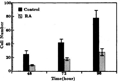
dUTP. Measurment of mRNA activity was carried out with conventional dot hybridization24. RESULTS INHIBITION OF IN VITRO INVASIVENESS OF CCL229 CELLS BY RA The number of cells which
penetrated the amnion in the group treated with RA (5 × 10⁻⁶ _M_) was 40 % of that of the control group at 48, 72, 96 h respectively indicating the inhibitive effects of RA on _in vitro_
invasiveness of CCL229 cells (P < 0.01) (Fig 1). EFFECTS OF RA ON THE MIGRATION OF CCL229 CELLS The migration of cancer cells was assessed by the number of cells which migrated out of the
agarose drops and the leading edge of the migration during a certain time interval. The number of cells that moved out of the drops in the group treated with RA were about half of those in
the control group (P < 0.01), and the leading edge in the experimental group was also significantly shorter than that in the controls (P <'0.01) (Tab 1). EFFECTS OF RA ON THE
ADHESIVENESS OF CCL229 CELLS The adhesiveness of the cancer cells was evaluated by the adhesion rate of the cells to the HABM. Fig 2 showed that the adhesiveness of CCL229 cells treated with
RA was near 30 % less than that of the controls (P < 0.01). EFFECTS OF RA ON SURFACE MORPHOLOGY OF CCL229 CELLS Scanning electron microscopy showed that CCL229 cells not treated with RA
assumed a fusiform shape with strong anchorage. Many microspikes and filopodia were revealed on the surface (Fig 3A). After treatment with RA, processes on the cell surface significantly
diminished, and the microspikes and filopodia virtually disappered (Fig 3B). EXPRESSION OF EGFR IN THE COLORECTAL CARCINOMA CELLS Dot hybridization with DIG labeled EGFR cDNA probe showed
that expression of EGFR in the highly invasive CCL229 cells was markedly higher than that in the low invasive CX-1 cells (Fig 4). The expression of EGFR in CCL229 cells treated with RA (5 ×
10−6 _M_) gradually decreased with time, and was hardly detectable on day 6 (Control not shown). DISCUSSION The primary factors which influence the invasiveness of cancer cells are their
migration, adhesiveness to the basement membrane and the capacity to secrete proteolytic enzymes to degrade the extracllular matrix25. Our previous study on the comparison of two well
differentiated cell lines: CCL229 and CX-1 cells showed that invasiveness, adhesiveness to the basement membrane, as well as the migration of CCL229 cells were significantly higher than
those of CX-1 cells, respectively. The morphology of the highly invasive CCL229 cells was characterized by numerous microspikes and filopodia on the cell surface26. These results indicated
that invasiveness of cancer cells was consistent with their adhesiveness and migration, and was correlated to the morphology of the cell surface. The present study demonstrated that 5 × 10⁻⁶
_M_ RA had obvious inhibitive effects on the _in vitro_ invasiveness, adhesiveness and migration of the CCL229 cells, and markedly reduced the microspikes and filopodia on the cell surface.
Although RA could affect the invasivehess through reducing the migration and adhesiveness to the basement membrane, the most crucial factor causing the penetration of the amnion rested on
the capacity of the cancer cells to secrete proteolytic enzymes that degraded the basement membrane and matrix. There have been reports that RA can inhibit the expression of collagenase and
stromelysin in some cancer cells9, 10, 11. These two are the most important proteolytic enzymes synthesized and secreted by the endoplasmic reticulum, and play a crucial role in the invasion
by cancer cells. Further investigations have shown that the promoters of the collagenase and stromelysin genes contain the binding sites for transcription factor AP1, and RA acts through
its nuclear receptor to reduce the activity of transcription factor AP1 and thus inhibit the expression of the two enzymes27. It was also reported that RA can inhibit the metastasis of human
breast carcinoma and invasion of rat mammary carcinoma to the basement membrane10, 12. There has been no published study, however, on the effects of RA on invasiveness of colorectal
carcinoma. The over-expression of EGFR in some cancers has received much attention. It appears that expression of EGFR might be used as a marker for prognosis. Salnsburg16 first noted that
expression of EGFR in highly invasive human breast carcinoma cells was higher than that in the benign tumors. Neal et al17 confirmed that EGFR was overexpressed in human invasive bladder
cancer cells. Yasui et al18 observed that EGFR in metastatic carcinomas in perigastric lymph nodes was markedly higher than that of primary carcinomas, and the expression of EGFR was
correlated to invasion, metastasis, and prognosis of gastric carcinoma. However, the relation between expression of EGFR and invasiveness of colorectal carcinoma remains obscure. The
observation that EGFR are differentially expressed in colorectal carcinoma cells with different degree of differentiation19 has prompted us to compare the EGFR expression of two cell lines
which are both well differentiated but have very different invasiveness. Our results showed that expression of EGFR in the very invasive CCL229 cells is significantly higher than that in the
low invasive CX-1 cells. In addition, expression of EGFR in CCL229 cells was down regulated as the invasiveness of CCL229 cells became decreased during the induction by × 10−6 _M_ RA. These
results all together indicate that invasiveness of colorectal carcinoma cells may be consistent with the degree of EGFR expression. There are similar reports demonstrating that RA may
inhibit expression of EGFR in some other cancer cells. Zheng et al28 found that RA inhibited EGFR expression in human epidermoid cancer cell line ME180 and that the inhibitive effect was
most evident when the concentration of RA was 10−5 M. They also showed that RA suppressed EGFR gene transcription and mRNA levels by its nuclear receptor. Another study reported that RA
decreased mRNA levels of EGFR by 2- to 4-folds in cultured human cerebrocervical squamous carcinoma cells 29. To our knowledge, our study is the first report on the inhibition by RA of
expression of EGFR in colorectal carcinoma cells. In conclusion, our study demonstrated that the expression of EGFR is higher in highly invasive colorectal carcinoma cells than that in low
invasive colorectal carcinoma cells; and the decrease of the expression of EGFR in a time- and dose-dependent manner after the cells are exposed to RA is accompanied by a synchronous
reduction of invasiveness of the caner cells. REFERENCES * Moon RC, McCormick DL, Mehta RG . Inhibition of carcinogenesis by retinoids. _Cancer Res_ 1983; 43: 2469–75. CAS Google Scholar *
Liu Youhua, Wang Yunqing . Studies on microscopic and submicroscopic structure of human promyelocytic leukemia cells (HL- 60) during differentiation induced by retinoids and dimethyl
sulfoxide. _Acta Biologiae Experimentalis Sinica_ 1985; 18(4): 389–404. Google Scholar * Lotan R . Effects of vitamin A and its anologs (retinoids) on normal and neoplastic cells. _Biochim
Biophys Acta_ 1980; 605: 33–91. CAS Google Scholar * Brown R . Retinoids alter the direction of differentiation in primary cultures of cutaneous keratinocytes. _Differentiation_ 1985; 28:
268. Article CAS Google Scholar * Sherman ME . Retinoids and Cell Differentiation. _Boca Raton FL: CRC Press Inc_ 1986. * Boyd AS . An overview of the retinoids. _Am J Med_
1989;86:568–574. Article CAS Google Scholar * Yao Zhaowei, Zha Xiliang, Ye Jiannan, Chen Huili . Some reverse effects of retinoic acid on the membranous phenotype of human hepatocarcinom
a cell line. _Chinese Journal of Biochemistry and Biophysics_ 1990; 22: 147–51. Google Scholar * Vasios GW, Gold JD, Patkovich M, Chambon P, Gudas LJ . A retinoic acid- responsive element
is present in the 5'flanking region of the laminin B1 gene. _Proc Natl Acad Sci USA_ 1989;86: 9099–103. Article CAS Google Scholar * Nakajima M, Lotan D, Baig M, Lotan R . Inhibition
by retinoic acid of type IV collagenolytic metalloproteinase production in metastatic rat mammary adenocarcinoma cells. _J Cell Biol_ 1987; 105(4, PART. 2):217a. Google Scholar * Nikajima
M, Lotan D, Baig MM, et al. Inhibition by retinoic acid of type IV collagenolysis and invasion through reconstituted basement membrane by metastatic rat mammary cancer. _Cancer Res_ 1989;49:
1698–706 . Google Scholar * Nicholson RC, Mader S, Nagpal S, Leid M, Rochette-Egly C, Chambon P . Negative regulation of the rat stromelysin gene promoter by retinoic acid is mediated by
an AP1 binding site. _EMBO J_ 1990; 9: 4443–54. Article CAS Google Scholar * Fraker LD, Halter SA, Forbes JT . Growth inhibition by retinol of a human breast carcinoma cell line in vitro
and in athymic mice. _Cancer Res_ 1984; 44: 5757–63. CAS PubMed Google Scholar * Hunts J, Ueda M, Ozawa S, Abe o, Pastan L, Shimizu N . Hyperproduction and gene amplification of the
epidermoid growth factor in squamous cell carcinomas. _Jpn J Cancer Res_ 1985; 76: 663–6. CAS PubMed Google Scholar * Veale D, Marsh C, Ashcroft T, Harris AL . Epidermal growth factor
receptors in non-small cell lung cancer. _Br J Cancer_ 1985; 52: 441. Google Scholar * Korc M, Meltzer P, Trent J . Enhanced expression of epidermal growth factor receptor correlates with
alternations of chromosome 7 in human pancreatic cancer. _Proc Natl Acad Sci USA_ 1986; 83: 5141–4. Article CAS Google Scholar * Salnsbury JRC, Farndon JR, Harris AL, Sherbet GV, Sherbet
GV . Epidermal growth factor receptors on human breast cancers. _Br J Surg_ 1985; 72: 186–8. Article Google Scholar * Neal DE, Marsh C . Epidermal growth factor receptors in human bladder
cancer: comparison of invasive and superficial tumours. _Lancet_ 1985; 1: 366–8. Article CAS Google Scholar * Yasui W, Sumiyoshi H, Hata J, et al. Expression of epidermal growth factor
receptor in human gastric and colonic carcinomas. _Cancer Res_ 1988; 48: 137–41. CAS PubMed Google Scholar * Bradley S J, Garfinkle G, Walker E, Salem R, Chen LB, Steel G . Increased
expression of the epidermal growth factor on human colon carcinoma cells. _Arch Surg_ 1986; 121: 1242–7. Article CAS Google Scholar * Liotta LA, Lee CW, Morakis DJ . New method for
preparing large surfaces of intact human basement membrane for tumor invasion studies. _Cancer Lett_ 1980; 11: 141–52. Article CAS Google Scholar * Varanl J, Orr W, Ward PA . A comparison
of the migration patterns of normal and malignant cells in two assay systems. _Am J Pathol_ 1978; 90: 159–71. Google Scholar * Murray JC, Liotta LA, Rennard SI, Martin GR . Adhesion
characteristics of murine metastatic and nonmetastatic tumor cells _in vitro_. _Cancer Res_ 1980; 40: 347–51. CAS PubMed Google Scholar * Chen Yuhua, Song Jindan . A simple, easy and
practical method for the preparation of RNA. Chinese Medical Biology Research. Cheng Du: Si Chuan Science and Technology Publishing House. 1995:235. * Sambrook J, Fritsch T, Maniatis T .
Molecular cloning. _A Laboratory manual. 2nd ed_. Cold Spring Harbor Laboratory Press 1989; 372–3. * Liotta LA . Cancer cell invasion and metastasis. _Scientific American_ 1992; February:
34–41. * Sun Baodong, Song Jindan . _In vitro_ comparative study on the invasive abiligy and its related biological properties of different colorectal caroinmoma cell lines. (to be
published). * Schule R, Rangarajan P, Yang N, et al. Retinoic acid is a negative regulator of AP-l-responsivegenes. _Proc Natl Acad Sci USA_ 1991; 88: 6092–6. Article CAS Google Scholar *
Zheng ZS, Goldsmith LA . Modulation of epidermal growth factor receptors by retinoic acid in ME180 cells. _Cancer Res_ 1990; 50:1201–5. CAS PubMed Google Scholar * Lei Wei, Wu Min .
Molecular mechanism of the regulation of retinoic acid in proliferation and differentiation. _Foreign Medical Molecular Biology: subunit_. 1994; 16(2): 83–6. Google Scholar Download
references AUTHOR INFORMATION AUTHORS AND AFFILIATIONS * Key Laboratory of Cell Biology, Ministry of Public Health of China, China Medical University, Shenyang, 110001, China Baodong Sun
& Jindan Song Authors * Baodong Sun View author publications You can also search for this author inPubMed Google Scholar * Jindan Song View author publications You can also search for
this author inPubMed Google Scholar ADDITIONAL INFORMATION *Dedicated to 80th anniversary of Professor Zhen YAO. †This work was supported by National Natural Science Foundation. RIGHTS AND
PERMISSIONS Reprints and permissions ABOUT THIS ARTICLE CITE THIS ARTICLE Sun, B., Song, J. Inhibition of invasiveness and expression of epidermal growth factor receptor in human colorectal
carcinoma cells induced by retinoic acid . _Cell Res_ 5, 135–142 (1995). https://doi.org/10.1038/cr.1995.13 Download citation * Received: 06 May 1995 * Revised: 12 May 1995 * Accepted: 14
May 1995 * Issue Date: 01 June 1995 * DOI: https://doi.org/10.1038/cr.1995.13 SHARE THIS ARTICLE Anyone you share the following link with will be able to read this content: Get shareable
link Sorry, a shareable link is not currently available for this article. Copy to clipboard Provided by the Springer Nature SharedIt content-sharing initiative KEYWORDS * Retinoic acid *
colorectal carcinoma cell * invasiveness * epidermal growth factor receptor * inhibition