Play all audios:
ABSTRACT Nanometric probes based on surface-enhanced Raman scattering (SERS) are promising candidates for all-optical environmental, biological and technological sensing applications with
intrinsic quantitative molecular specificity. However, the effectiveness of SERS probes depends on a delicate trade-off between particle size, stability and brightness that has so far
hindered their wide application in SERS imaging methodologies. In this Article, we introduce holographic Raman microscopy, which allows single-shot three-dimensional single-particle
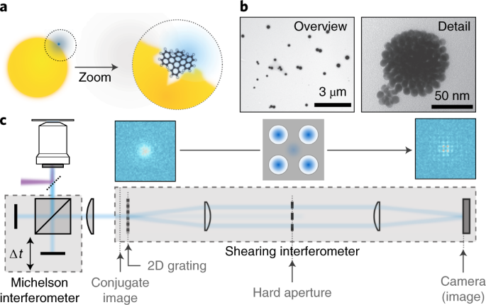
localization. We validate our approach by simultaneously performing Fourier transform Raman spectroscopy of individual SERS nanoparticles and Raman holography, using shearing interferometry
to extract both the phase and the amplitude of wide-field Raman images and ultimately localize and track single SERS nanoparticles inside living cells in three dimensions. Our results
represent a step towards multiplexed single-shot three-dimensional concentration mapping in many different scenarios, including live cell and tissue interrogation and complex
anti-counterfeiting applications. Access through your institution Buy or subscribe This is a preview of subscription content, access via your institution ACCESS OPTIONS Access through your
institution Access Nature and 54 other Nature Portfolio journals Get Nature+, our best-value online-access subscription $29.99 / 30 days cancel any time Learn more Subscribe to this journal
Receive 12 print issues and online access $259.00 per year only $21.58 per issue Learn more Buy this article * Purchase on SpringerLink * Instant access to full article PDF Buy now Prices
may be subject to local taxes which are calculated during checkout ADDITIONAL ACCESS OPTIONS: * Log in * Learn about institutional subscriptions * Read our FAQs * Contact customer support
SIMILAR CONTENT BEING VIEWED BY OTHERS INTERFEROMETRIC SCATTERING MICROSCOPY Article 10 April 2025 DETECTION OF SURFACE ENHANCED RAMAN SCATTERING ACTIVE HOTSPOT USING NEAR FIELD SCANNING
OPTICAL MICROSCOPY Article Open access 08 May 2024 SINGLE-SHOT ISOTROPIC DIFFERENTIAL INTERFERENCE CONTRAST MICROSCOPY Article Open access 12 April 2023 DATA AVAILABILITY The materials and
data that support the findings of this study are available from the corresponding authors on request. CODE AVAILABILITY The software used for data analysis is available from the
corresponding authors on request. REFERENCES * Albrecht, M. G. & Creighton, J. A. Anomalously intense Raman spectra of pyridine at a silver electrode. _J. Am. Chem. Soc._ 4009, 5215–5217
(1977). Article Google Scholar * Jeanmaire, D. L. & Van Duyne, R. P. Surface Raman spectroelectrochemistry: Part 1. Heterocyclic, Aromatic, and aliphatic amines adsorbed on the
anodized silver electrode. _J. Electroanal. Chem. Interfacial Electrochem._ 84, 1–20 (1977). Article CAS Google Scholar * Fleischmann, M., Hendra, P. J. & McQuillan, A. J. Raman
spectra of pyridine absorbed at a silver electrode. _Chem. Phys. Lett._ 26, 163–166 (1974). Article CAS Google Scholar * Langer, J. et al. Present and future of surface-enhanced Raman
scattering. _ACS Nano_ 14, 28–117 (2020). Article CAS Google Scholar * Lane, L. A., Qian, X. & Nie, S. SERS nanoparticles in medicine: from label-free detection to spectroscopic
tagging. _Chem. Rev._ 115, 10489–10529 (2015). Article CAS Google Scholar * Wang, Y., Yan, B. & Chen, L. SERS tags: novel optical nanoprobes for bioanalysis. _Chem. Rev._ 113,
1391–1428 (2013). Article CAS Google Scholar * Zhang, X., Young, M. A., Lyandres, O. & Van Duyne, R. P. Rapid detection of an anthrax biomarker by surface-enhanced Raman spectroscopy.
_J. Am. Chem. Soc._ 127, 4484–4489 (2005). Article CAS Google Scholar * Grubisha, D. S., Lipert, R. J., Park, H.-Y., Driskell, J. & Porter, M. D. Femtomolar detection of
prostate-specific antigen: an immunoassay based on surface-enhanced Raman scattering and immunogold labels. _Anal. Chem._ 75, 5936–5943 (2003). Article CAS Google Scholar * Pazos, E. et
al. Surface-enhanced Raman scattering surface selection rules for the proteomic liquid biopsy in real samples: efficient detection of the oncoprotein c-MYC. _J. Am. Chem. Soc._ 138,
14206–14209 (2016). Article CAS Google Scholar * Li, J. F. et al. Shell-isolated nanoparticle-enhanced Raman spectroscopy. _Nature_ 464, 392–395 (2010). Article CAS Google Scholar *
Dasary, S. S. R., Singh, A. K., Senapati, D., Yu, H. & Ray, P. C. Gold nanoparticle based label-free SERS probe for ultrasensitive and selective detection of trinitrotoluene. _J. Am.
Chem. Soc._ 131, 13806–13812 (2009). Article CAS Google Scholar * Pazos-Perez, N. et al. Ultrasensitive multiplex optical quantification of bacteria in large samples of biofluids. _Sci.
Rep._ 6, 29014 (2016). Article CAS Google Scholar * Pallaoro, A., Hoonejani, M. R., Braun, G. B., Meinhart, C. D. & Moskovits, M. Rapid identification by surface-enhanced Raman
spectroscopy of cancer cells at low concentrations flowing in a microfluidic channel. _ACS Nano_ 9, 4328–4336 (2015). Article CAS Google Scholar * Palonpon, A. F. et al. Raman and SERS
microscopy for molecular imaging of live cells. _Nat. Protoc._ 8, 677–692 (2013). Article CAS Google Scholar * Rivera-Gil, P. et al. Plasmonic nanoprobes for real-time optical monitoring
of nitric oxide inside living cells. _Angew. Chem. Int. Ed._ 125, 13939–13943 (2013). Article Google Scholar * Phan-Quang, G. C. et al. Three-dimensional surface-enhanced Raman scattering
platforms: large-scale plasmonic hotspots for new applications in sensing, microreaction, and data storage. _Acc. Chem. Res._ 52, 1844–1854 (2019). Article CAS Google Scholar * Jiang, W.,
Kim, B. Y. S., Rutka, J. T. & Chan, W. C. W. Nanoparticle-mediated cellular response is size-dependent. _Nat. Nanotechnol._ 3, 145–150 (2008). Article CAS Google Scholar *
Latychevskaia, T. & Fink, H.-W. Holographic time-resolved particle tracking by means of three-dimensional volumetric deconvolution. _Opt. Express_ 22, 20994–21003 (2014). Article Google
Scholar * Molaei, M. & Sheng, J. Imaging bacterial 3D motion using digital in-line holographic microscopy and correlation-based de-noising algorithm. _Opt. Express_ 22, 32119–32137
(2014). Article Google Scholar * Memmolo, P. et al. Recent advances in holographic 3D particle tracking. _Adv. Opt. Photonics_ 7, 713–755 (2015). Article Google Scholar * Gabor, D. A new
microscopic principle. _Nature_ 161, 777–778 (1948). Article CAS Google Scholar * Cuche, E., Marquet, P. & Depeursinge, C. Spatial filtering for zero-order and twin-image elimination
in digital off-axis holography. _Appl. Opt._ 39, 4070–4075 (2000). Article CAS Google Scholar * Bon, P. et al. Self-interference 3D super-resolution microscopy for deep tissue
investigations. _Nat. Methods_ 15, 449–454 (2018). Article CAS Google Scholar * Patorski, K. The self-imaging phenomenon and its applications. _Prog. Opt._ 27, 1–108 (1989). Article
Google Scholar * Bon, P., Maucort, G., Wattellier, B. & Monneret, S. Quadriwave lateral shearing interferometry for quantitative phase microscopy of living cells. _Opt. Express_ 17,
13080–13094 (2009). Article CAS Google Scholar * Camden, J. P. et al. Probing the structure of single-molecule surface-enhanced Raman scattering hot spots. _J. Am. Chem. Soc._ 130,
12616–12617 (2008). Article CAS Google Scholar * Michaels, A. M., Jiang, J. & Brus, L. Ag nanocrystal junctions as the site for surface-enhanced Raman scattering of single rhodamine
6G molecules. _J. Phys. Chem. B_ 104, 11965–11971 (2000). Article CAS Google Scholar * Maznev, A. A., Crimmins, T. F. & Nelson, K. A. How to make femtosecond pulses overlap. _Opt.
Lett._ 23, 1378–1380 (1998). Article CAS Google Scholar * Takeda, M., Ina, H. & Kobayashi, S. Fourier-transform method of fringe-pattern analysis for computer-based topography and
interferometry. _J. Opt. Soc. Am._ 72, 156–160 (1982). Article Google Scholar * Arnison, M. R., Larkin, K. G., Sheppard, C. J. R., Smith, N. I. & Cogswell, C. J. Linear phase imaging
using differential interference contrast microscopy. _J. Microsc._ 214, 7–12 (2003). Article Google Scholar * Choi, I., Lee, K. & Park, Y. Compensation of aberration in quantitative
phase imaging using lateral shifting and spiral phase integration. _Opt. Express_ 25, 30771–30779 (2017). Article Google Scholar * Jaqaman, K. et al. Robust single-particle tracking in
live-cell time-lapse sequences. _Nat. Methods_ 5, 695–702 (2008). Article CAS Google Scholar * Rubin, M., Dardikman, G., Mirsky, S. K., Turko, N. A. & Shaked, N. T. Six-pack off-axis
holography. _Opt. Lett._ 42, 4611–4614 (2017). Article Google Scholar Download references ACKNOWLEDGEMENTS We thank M. Rivas for her support with the live-cell experiments. We acknowledge
support by the Ministry of Science, Innovation and Universities (MCIU/AEI: RTI2018-099957-J-I00 and PGC2018-096875-B-I00), the Ministry of Economy (MINECO: CTQ2017-88648-R, RYC-2015-19107
and ‘Severo Ochoa’ programme for Centers of Excellence in R&D CEX2019-000910-S), the Catalan AGAUR (2017SGR1369 and 2017SGR883), Fundació Privada Cellex, Fundació Privada Mir-Puig, the
Generalitat de Catalunya through the CERCA programme and the Universitat Rovira i Virgili (FR 2019-B2). N.F.v.H. acknowledges the financial support by the European Commission (ERC Advanced
Grant 670949-LightNet). AUTHOR INFORMATION AUTHORS AND AFFILIATIONS * ICFO — Institut de Ciencies Fotoniques, The Barcelona Institute of Science and Technology, Barcelona, Spain Matz Liebel
& Niek F. van Hulst * Department of Physical and Inorganic Chemistry and EMaS, Universitat Rovira i Virgili, Tarragona, Spain Nicolas Pazos-Perez & Ramon A. Alvarez-Puebla * ICREA —
Institució Catalana de Recerca i Estudis Avançats, Barcelona, Spain Niek F. van Hulst & Ramon A. Alvarez-Puebla Authors * Matz Liebel View author publications You can also search for
this author inPubMed Google Scholar * Nicolas Pazos-Perez View author publications You can also search for this author inPubMed Google Scholar * Niek F. van Hulst View author publications
You can also search for this author inPubMed Google Scholar * Ramon A. Alvarez-Puebla View author publications You can also search for this author inPubMed Google Scholar CONTRIBUTIONS M.L.
constructed the optical experiment, performed the measurements and analysed the data. M.L. conceived the experiment. N.P.P. and R.A.A.P. synthesized and characterized the nanoparticles. M.L.
wrote the manuscript. M.L., N.F.v.H. and R.A.A.P. contributed to the interpretation of the data, discussion and writing of the manuscript. CORRESPONDING AUTHORS Correspondence to Matz
Liebel, Niek F. van Hulst or Ramon A. Alvarez-Puebla. ETHICS DECLARATIONS COMPETING INTERESTS The authors declare no competing interests. ADDITIONAL INFORMATION PEER REVIEW INFORMATION
_Nature Nanotechnology_ thanks Pasquale Memmolo and the other, anonymous, reviewer(s) for their contribution to the peer review of this work. Peer reviewer reports are available. PUBLISHER’S
NOTE Springer Nature remains neutral with regard to jurisdictional claims in published maps and institutional affiliations. SUPPLEMENTARY INFORMATION SUPPLEMENTARY INFORMATION Supplementary
Figs. 1–8 and Information S1–S7. SUPPLEMENTARY VIDEO 1 Live-cell tracking video supporting Fig. 6, showing bright-field (grey) and SERS (pink) signals. RIGHTS AND PERMISSIONS Reprints and
permissions ABOUT THIS ARTICLE CITE THIS ARTICLE Liebel, M., Pazos-Perez, N., van Hulst, N.F. _et al._ Surface-enhanced Raman scattering holography. _Nat. Nanotechnol._ 15, 1005–1011 (2020).
https://doi.org/10.1038/s41565-020-0771-9 Download citation * Received: 20 March 2020 * Accepted: 26 August 2020 * Published: 28 September 2020 * Issue Date: December 2020 * DOI:
https://doi.org/10.1038/s41565-020-0771-9 SHARE THIS ARTICLE Anyone you share the following link with will be able to read this content: Get shareable link Sorry, a shareable link is not
currently available for this article. Copy to clipboard Provided by the Springer Nature SharedIt content-sharing initiative